. 2008 May 2;283(18):12166-74.
doi: 10.1074/jbc.M800763200. Epub 2008 Feb 29.

Molecular anatomy of the recombination mediator function of Saccharomyces cerevisiae Rad52

Affiliations

Molecular anatomy of the recombination mediator function of Saccharomyces cerevisiae Rad52

Changhyun Seong et al. J Biol Chem. .

Abstract

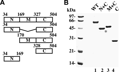
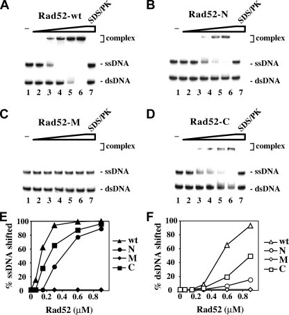
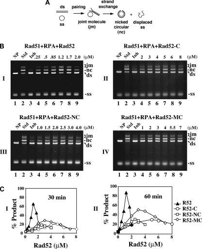
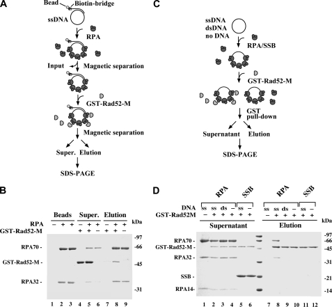
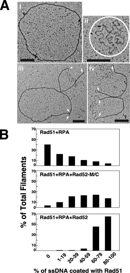
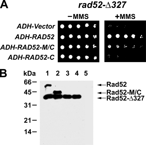
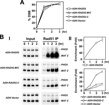
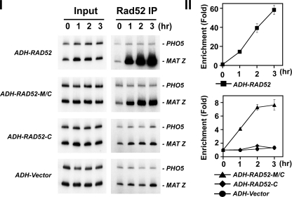
A helical filament of Rad51 on single-strand DNA (ssDNA), called the presynaptic filament, catalyzes DNA joint formation during homologous recombination. Rad52 facilitates presynaptic filament assembly, and this recombination mediator activity is thought to rely on the interactions of Rad52 with Rad51, the ssDNA-binding protein RPA, and ssDNA. The N-terminal region of Rad52, which has DNA binding activity and an oligomeric structure, is thought to be crucial for mediator activity and recombination. Unexpectedly, we find that the C-terminal region of Rad52 also harbors a DNA binding function. Importantly, the Rad52 C-terminal portion alone can promote Rad51 presynaptic filament assembly. The middle portion of Rad52 associates with DNA-bound RPA and contributes to the recombination mediator activity. Accordingly, expression of a protein species that harbors the middle and C-terminal regions of Rad52 in the rad52 Delta327 background enhances the association of Rad51 protein with a HO-made DNA double-strand break and partially complements the methylmethane sulfonate sensitivity of the mutant cells. Our results provide a mechanistic framework for rationalizing the multi-faceted role of Rad52 in recombination and DNA repair.


Figures

FIGURE 1.
FIGURE 1.
Purification of Rad52 species. A, schematic representation of the Rad52 species used. B, purified His6-tagged proteins: Rad52, Rad52-N/C, Rad52-M/C, and Rad52-C (1 μg each) were run on a 10% denaturing SDS-PAGE and stained with Coomassie Blue. The asterisk designates a proteolytic fragment of Rad52-N/C.
FIGURE 2.
FIGURE 2.
The C-terminal region of Rad52 harbors a DNA binding activity. A–D, Radiolabeled ssDNA and dsDNA were incubated without protein (lane 1) and with 60, 135, 270, and 400 nm of Rad52 (A), Rad52-N (B), Rad52-M (C), and Rad52-C (D). In lane 7, a reaction mixture containing the highest amount of each of the Rad52 species was treated with SDS and proteinase K before analysis. The results from A–D were plotted in E and F. Note that the ssDNA migrated more slowly than the dsDNA. wt, wild type.
FIGURE 3.
FIGURE 3.
The C terminus of Rad52 possesses recombination mediator activity. A, schematic representation of the homologous DNA pairing and strand exchange reaction. Homologous pairing between the circular ssDNA (ss) and linear duplex DNA (ds) substrates yields a DNA joint molecule (jm), which is converted into a nicked circular duplex molecule (nc) by DNA strand exchange. B, DNA strand exchange reactions conducted to examine the recombination mediator activity of the Rad52 species: Rad52 in panel I, Rad52-C in panel II, Rad52-N/C in panel III, and Rad52-M/C in panel IV. The standard reaction (Std, lane 2) involved preincubating the ssDNA with Rad51 to allow for the formation of the presynaptic filament before RPA was added. Co-incubating the ssDNA with Rad51 and RPA resulted in severe inhibition of the DNA strand exchange reaction (Inh, lane 3). The inclusion of the various Rad52 species during the incubation of ssDNA with Rad51 and RPA led to restoration of DNA strand exchange. NP indicates no protein added. C, the results of the 30- and 60-min time points in B were plotted in panels I and II, respectively.
FIGURE 4.
FIGURE 4.
Rad52-M interacts with DNA-bound RPA. A, in this affinity pulldown assay, RPA is incubated with streptavidin magnetic beads harboring φX174 ssDNA, and the resulting RPA-containing beads are used to bind Rad52-M. B, three reactions were set up. In the first reaction, ssDNA magnetic beads that did not harbor RPA (lane 1) were mixed with GST-Rad52-M (lanes 4 and 7); in the second reaction, ssDNA magnetic beads harboring RPA (lane 2) were mixed with GST-Rad52-M (lanes 5 and 8); and in the third reaction, ssDNA magnetic beads harboring RPA (lane 3) were mixed in buffer without GST-Rad52-M (lanes 6 and 9). After mixing, the beads were captured with a magnet, and associated proteins were eluted with SDS. The supernatants (Super.) that contained unbound proteins and the SDS eluates (Elution) were analyzed by SDS-PAGE with Coomassie Blue staining. The RPA content of the magnetic beads (Beads) used in the three reactions is also shown. C, in this affinity pulldown assay, GST-Rad52-M is incubated with RPA or E. coli SSB either in the absence of DNA or in the presence of ssDNA or dsDNA before being mixed with glutathione-Sepharose to capture GST-Rad52-M and any associated RPA. D, the various supernatants containing unbound proteins and the SDS eluates harboring the captured proteins were analyzed by SDS-PAGE with Coomassie Blue staining. GST-Rad52-M was omitted from the control reaction in lane 1.
FIGURE 5.
FIGURE 5.
Electron microscopy analysis of recombination mediator action. A, micrographs showing an example of full Rad51 filament (panel i), a RPA-ssDNA nucleoprotein complex (panel ii), and ssDNA molecules covered partly by Rad51 and partly by RPA (panels iii and iv). The RPA-ssDNA complexes in panel ii are circled, and the arrows in panels iii and iv mark the junctions of Rad51-coated DNA and RPA-coated DNA. The black scale bars in the panels denote 200 nm. B, quantification of Rad51 presynaptic filament formation in the three reactions: Rad51+RPA, Rad51+RPA+ Rad52-M/C, and Rad51+RPA+Rad52.
FIGURE 6.
FIGURE 6.
Complementation of the rad52-Δ327 mutant by Rad52-M/C. A, rad52-Δ327 strains harboring the empty ADH vector, ADH-RAD52, ADH-RAD52-M/C, or ADH-RAD52-C were serially diluted and spotted onto SC-Ura medium with or without 0. 005% MMS. B, extracts of rad52-Δ327 cells harboring ADH plasmids expressing Rad52 (lane 1), Rad52-M/C (lane 2), Rad52-C (lane 3), or empty ADH vector (lane 4) or rad52Δ cells harboring empty ADH vector (lane 5) were subjected to immunoblot analysis with anti-Rad52 antibodies.
FIGURE 7.
FIGURE 7.
Rad52-M/C promotes DSB recruitment of Rad51. A, kinetics of DSB induction. Donorless rad52-Δ327 cells harboring GAL10-HO and also the empty ADH vector or a ADH plasmid expressing Rad52, Rad52-M/C, or Rad52-C were grown in the presence of galactose to induce HO-expression and a DSB at MAT. The induction of the HO break was quantified by PCR (20, 21). B, recruitment of Rad51 to the HO-induced break in rad52-Δ327 cells harboring the ADH vector or ADH-RAD52, ADH-RAD52-M/C, or RAD52-C. These cells were subjected to ChIP with anti-Rad51 antibodies. The MAT Z target sequence and control PHO5 sequence were amplified using the appropriate primer sets (21). Panels I and II show the PCR products and the quantification of the results, respectively. Enrichment was determined by dividing the PCR signal of MAT Z by the PHO5 PCR signal and normalizing to time zero value.
FIGURE 8.
FIGURE 8.
DSB recruitment of Rad52 species in rad52-Δ327 cells. Recruitment of Rad52 to the HO breaks in rad52-Δ327 cells harboring the ADH vector or ADH-RAD52, ADH-RAD52-M/C, or ADH-RAD52-C. These cells were subjected to ChIP with anti-Rad52 antibodies. The MAT Z target sequence and control PHO5 sequence were amplified using the appropriate primer sets (21). Panels I and II show the PCR products and the quantification of the results, respectively. Enrichment was determined by dividing the PCR signal of MAT Z by the PHO5 PCR signal and normalizing to time zero value.

References

    1. Cox, M. M., Goodman, M. F., Kreuzer, K. N., Sherratt, D. J., Sandler, S. J., and Marians, K. J. (2000) Nature 404 37–41 - PubMed
    1. Symington, L. S. (2002) Microbiol. Mol. Biol. Rev. 66 630–670 - PMC - PubMed
    1. Heller, R. C., and Marians, K. J. (2006) Nat. Rev. Mol. Cell Biol. 7 932–943 - PubMed
    1. Jasin, M. (2002) Oncogene 21 8981–8993 - PubMed
    1. D'Andrea, A. D. (2003) Genes Dev. 17 1933–1936 - PubMed

Publication types

MeSH terms