. 2008 May;29(5):1005-12.
doi: 10.1093/carcin/bgn007. Epub 2008 Feb 28.

Superior in vivo inhibitory efficacy of methylseleninic acid against human prostate cancer over selenomethionine or selenite

Affiliations

Superior in vivo inhibitory efficacy of methylseleninic acid against human prostate cancer over selenomethionine or selenite

Guang-xun Li et al. Carcinogenesis. 2008 May.

Abstract

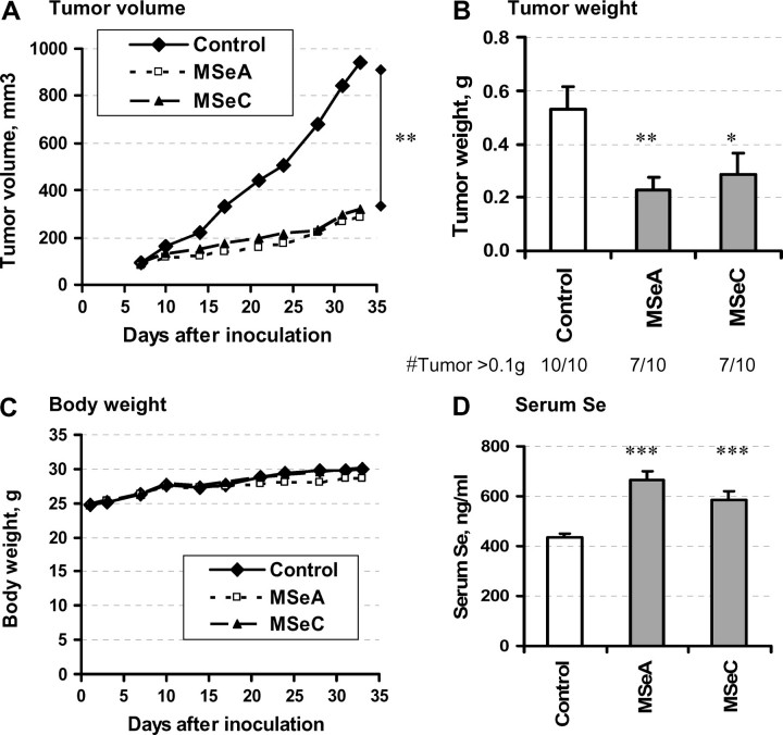
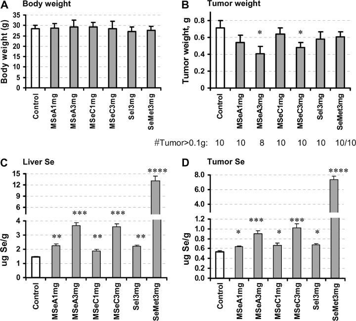
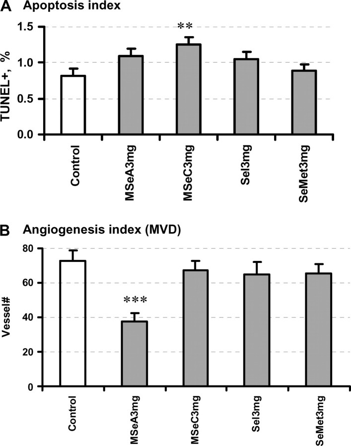
Methylselenol has been implicated as an active anticancer selenium (Se) metabolite. However, its in vivo efficacy against prostate cancer (PCa) has yet to be established. Here, we evaluated the growth inhibitory effects of two presumed methylselenol precursors methylseleninic acid (MSeA) and Se-methylselenocysteine (MSeC) in comparison with selenomethionine (SeMet) and selenite in DU145 and PC-3 human PCa xenografts in athymic nude mice. Each Se was given by a daily single oral dose regimen starting the day after the subcutaneous inoculation of cancer cells. We analyzed serum, liver and tumor Se content to confirm supplementation status and apoptosis indices and tumor microvessel density for association with antitumor efficacy. Furthermore, we analyzed lymphocyte DNA integrity to detect genotoxic effect of Se treatments. The data show that MSeA and MSeC exerted a dose-dependent inhibition of DU145 xenograft growth and both were more potent than SeMet and selenite, in spite of less tumor Se retention than in the SeMet-treated mice. Selenite treatment increased DNA single-strand breaks in peripheral lymphocytes, whereas the other Se forms did not. Terminal deoxynucleotidyl transferase biotin-dUTP nick end labeling (TUNEL) and cleaved caspase-3 indices (apoptosis) from MSeC-treated tumors were higher than tumors from control mice or MSeA-treated mice, whereas the microvessel density index was lower in tumors from MSeA-treated mice. In the PC-3 xenograft model, only MSeA was growth inhibitory at a dose of 3 mg/kg body wt. In summary, our data demonstrated superior in vivo growth inhibitory efficacy of MSeA over SeMet and selenite, against two human PCa xenograft models without the genotoxic property of selenite.


Figures

Fig. 1.
Fig. 1.
Effects of a daily single oral dose of MSeA and MSeC on the growth of DU145 PCa subcutaneous xenografts in athymic nude mice. Dosage for MSeA and MSeC was 4 mg Se/kg body wt. (A) Mean tumor volume as a function of time after inoculation. (B) Final tumor weight after necropsy, Mean ± SE, n = 10 mice. (C) Mean body weight of mice during the course of experiment. (D) Serum Se content at 24 h after the last Se dose, mean ± SE, n = 10 mice. Statistical significance from control group: *P < 0.05 and **P < 0.01. Experiment 1.
Fig. 2.
Fig. 2.
Comparison of effects of MSeA and MSeC on apoptosis and angiogenesis indices. (A) TUNEL and (B) cleaved caspase-3 as biomarkers of cancer epithelial cell apoptosis. Mean ± SE, n = 7–10. Statistical significance from control group: *P < 0.05. (C) Microvessel counts as a biomarker of tumor angiogenesis, Mean ± SE, n = 7–10. Statistical significance from control group: **P <0.01. Experiment 1.
Fig. 3.
Fig. 3.
The effects of a daily single oral dose of MSeA and MSeC in comparison with sodium selenite (Sel) and SeMet on the growth of DU145 PCa subcutaneous xenografts in athymic nude mice and their liver and tumor Se contents. (A) Body weight of mice at termination of experiment (49 days). Mean ± SD. Not statistical significant. (B) Tumor weight after necropsy. Mean ± SE, n = 10. (C) Liver Se content. Mean ± SE, n = 10. (D) Xenograft tumor Se content. Mean ± SE, n = 8–10. Statistical significance from control group: *P < 0.05, **P < 0.01, ***P < 0.001 and ****P < 0.0001. Experiment 2.
Fig. 4.
Fig. 4.
Evaluation of (A) TUNEL index for DU145 xenograft cancer epithelial apoptosis and (B) microvessel counts as a biomarker of angiogenesis. Se dose level was 3 mg Se/kg body wt. Mean ± SE, n = 8–10. Statistical significance from control group: **P <0.01 and ***P < 0.001. Experiment 2.
Fig. 5.
Fig. 5.
Assessment of DNA integrity by single cell electrophoresis assay (COMET) of the peripheral lymphocytes of athymic nude mice after 48 days of daily oral treatment with MSeA, MSeC, selenite and SeMet. (A) Representative images of lymphocyte nuclei from a control mouse (left panel) and from a selenite-treated mouse (right panel). The fantails indicate DNA strand breakage. (B) Proportion of nuclei with moderate-to-extensive DNA strand breaks under alkaline electrophoresis condition (measuring both SSB and double-strand breaks). A total of 100 nuclei were scored for each mouse. (C) Proportion of nuclei with moderate-to-extensive DNA strand breaks under neutral electrophoresis condition (measuring double-strand breaks). A total of 200 nuclei were scored for each mouse. Mean ± SE, n = 10 mice. Statistical significance from control group: ***P < 0.001. Experiment 2.
Fig. 6.
Fig. 6.
The effects of a daily single oral dose of MSeA, MSeC, sodium selenite and SeMet on the growth of PC-3 PCa subcutaneous xenografts in athymic nude mice. (A) Body weight of mice before termination of experiment. Mean ± SD, n = 12–16. (B) Tumor weight after necropsy. Mean ± SE, n = 16. Statistical significance from control group: *P < 0.05 (one sided). Euthanasia # reflects the number of tumor-bearing mice requiring euthanasia due to heavy tumor burden to comply with institutional animal care and use committee regulation before the preset termination time. (C) Rank-order plot of individual PC-3 tumors from the five groups from heaviest to lightest. Experiment 3.

References

    1. Jemal A, et al. Cancer statistics, 2006. CA Cancer J. Clin. 2006;56:106–130. - PubMed
    1. Petrylak DP. The current role of chemotherapy in metastatic hormone-refractory prostate cancer. Urology. 2005;65:3–7. discussion 7–8. - PubMed
    1. Clark LC, et al. Effects of selenium supplementation for cancer prevention in patients with carcinoma of the skin. A randomized controlled trial. Nutritional Prevention of Cancer Study Group. JAMA. 1996;276:1957–1963. - PubMed
    1. Lippman SM, et al. Designing the Selenium and Vitamin E Cancer Prevention Trial (SELECT) J. Natl Cancer Inst. 2005;97:94–102. - PubMed
    1. Ip C. Lessons from basic research in selenium and cancer prevention. J. Nutr. 1998;128:1845–1854. - PubMed

Publication types