. 2008 Mar 11;105(10):3663-7.
doi: 10.1073/pnas.0707223105. Epub 2008 Feb 29.

Microfluidics of cytoplasmic streaming and its implications for intracellular transport

Affiliations

Microfluidics of cytoplasmic streaming and its implications for intracellular transport

Raymond E Goldstein et al. Proc Natl Acad Sci U S A. .

Abstract

Found in many large eukaryotic cells, particularly in plants, cytoplasmic streaming is the circulation of their contents driven by fluid entrainment from particles carried by molecular motors at the cell periphery. In the more than two centuries since its discovery, streaming has frequently been conjectured to aid in transport and mixing of molecular species in the cytoplasm and, by implication, in cellular homeostasis, yet no theoretical analysis has been presented to quantify these processes. We show by a solution to the coupled dynamics of fluid flow and diffusion appropriate to the archetypal "rotational streaming" of algal species such as Chara and Nitella that internal mixing and the transient dynamical response to changing external conditions can indeed be enhanced by streaming, but to an extent that depends strongly on the pitch of the helical flow. The possibility that this may have a developmental consequence is illustrated by the coincidence of the exponential growth phase of Nitella and the point of maximum enhancement of those processes.


Conflict of interest statement

The authors declare no conflict of interest.

Figures

Fig. 1.
Fig. 1.
Architecture of Chara corallina. (a) Portion of a plant, with leaf cells (1), nodes (2), and internodal cells (3). (b) Enlargement of an internodal cell, displaying spiraling indifferent zone. (c) Close-up of the indifferent zone. (d) Schematic cross-section: chloroplasts (4), indifferent zone (5), endoplasm (6), and vacuole (7).
Fig. 2.
Fig. 2.
Idealized spiraling flow in Chara. (a) Flow at the boundary, divided in an ascending band (red) and descending band (blue) separated by two indifferent zones (yellow, labeled IZ+/−). Vectors indicate the direction of flow along the bands. Arrows labeled H and ϕ indicate the axes parallel and perpendicular to the spiral. The shaded region corresponds to the horizontal section shown by its intersection with the boundary as a horizontal solid line in b and viewed along the cell axis in c and d. (b) Cylinder from a, cut open along dotted line (as indicated by scissors) and flattened out. Ascending and descending regions now appear as diagonal bands. The two indifferent zones have a subtle difference in symmetry, which is reflected in the horizontal components of motion converging at one zone and diverging for the other, as indicated by the arrows at bottom. (c) Projection of the flow along the spiraling axis (with z axis into page), for a cross-section marked by the circle in a, with λ/R = 3. Contours and arrows denote the z− and xy− components of v(r, ϕ)H, respectively. (d) Velocity field components orthogonal to H, denoted by J(r, ϕ). This transversal flow is smaller than the spiraling component by an order of magnitude and is zero at the wall, where flow is parallel to the spiral.
Fig. 3.
Fig. 3.
Spiralling flow. (a) Trajectory of a fluid parcel over one cycle of longitudinal transport for λ/R = 3. (b) Projection of the trajectory onto the xy− plane.
Fig. 4.
Fig. 4.
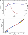
Wavelength dependence and comparison with experiments. (a) Root-mean-square longitudinal (blue) and transversal (red) velocities as functions of helical wavelength. (b) Comparison of solved velocity to experimental data extracted from Kamiya and Kuroda (7) (yellow squares) and Mustacich (32) (blue triangles), where the velocities have been normalized to their maximum values, at the cell surface.
Fig. 5.
Fig. 5.
Time-dependence of concentration during diffusion into cell. (a) In a cell with straight indifferent zones the concentration in a cross-section is azimuthally symmetric. (b) With a finite pitch (λ/R = 3) and Pe = 500 advection through the center of the cell (Fig. 2d) redistributes material, resulting in a steep concentration gradient at the left indifferent zone, which leads to a significant increase in the flux across the boundary. (c and d) Concentration at the center of the cell (c) and flux into a small cylinder at the center (d), for a series of Péclet numbers 10, 20, 50, …, 104. Low Péclet numbers are colored blue, and high Péclet numbers are colored red. Diffusion into the cell is significantly enhanced for high Pe.
Fig. 6.
Fig. 6.
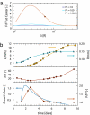
Wavelength variations. (a) Flux enhancement at the center of a cell with constant concentration boundary conditions at the cell wall as a function of helix wavelength, at various Pe. (b) Comparison of calculated flux with data on growth of Nitella axillaris Braun, from Green (36). Blue triangles, cell diameter; orange squares, cell length; orange circles, helical wavelength in units of the cell radius. The flux enhancement (orange circles) at the cell center is calculated from model with rotational streaming and the nondimensionalized surface growth rate S−1dS/dt (blue solid curve), where S is the total surface area, calculated from spline-interpolated diameter and length. Arrowheads in a show the trajectory of wavelength over time, first decreasing and then increasing.
Fig. 7.
Fig. 7.
Heterogeneity in the uptake rate of a cell for λ/R = 12. (a) Color-coded rate of uptake on the cell surface, for Pe = 1,000. (b) Enhancement of flux through a circular region near the cell wall due to fluid flow for various values of the Péclet number (0, 10, 20, 50, …, 103), as a function of azimuthal angle.

Similar articles

Cited by

References

    1. Corti B. Osservazione Microscopische sulla Tremella e sulla Circulazione del Fluido in Una Planto Acquaguola. Lucca, Italy: Appresso Giuseppe Rocchi; 1774.
    1. Pickard WF. Plant Cell Environ. 2003;26:1–15.
    1. Allen RD, Allen NS. Annu Rev Biophys Bioeng. 1978;7:469–495. - PubMed
    1. Shimmen T. J Plant Res. 2007;120:31–43. - PubMed
    1. Allen NS, Allen RD. Annu Rev Biophys Bioeng. 1978;7:497–526. - PubMed

Publication types

LinkOut - more resources