. 2008 Apr;172(4):1100-11.
doi: 10.2353/ajpath.2008.070457. Epub 2008 Feb 29.

Human immunodeficiency virus (HIV) infects human arterial smooth muscle cells in vivo and in vitro: implications for the pathogenesis of HIV-mediated vascular disease

Affiliations

Human immunodeficiency virus (HIV) infects human arterial smooth muscle cells in vivo and in vitro: implications for the pathogenesis of HIV-mediated vascular disease

Eliseo A Eugenin et al. Am J Pathol. 2008 Apr.

Abstract

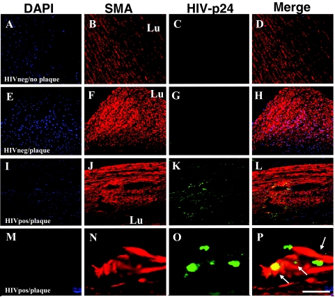
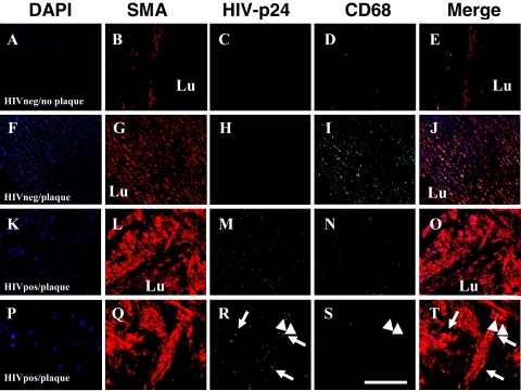
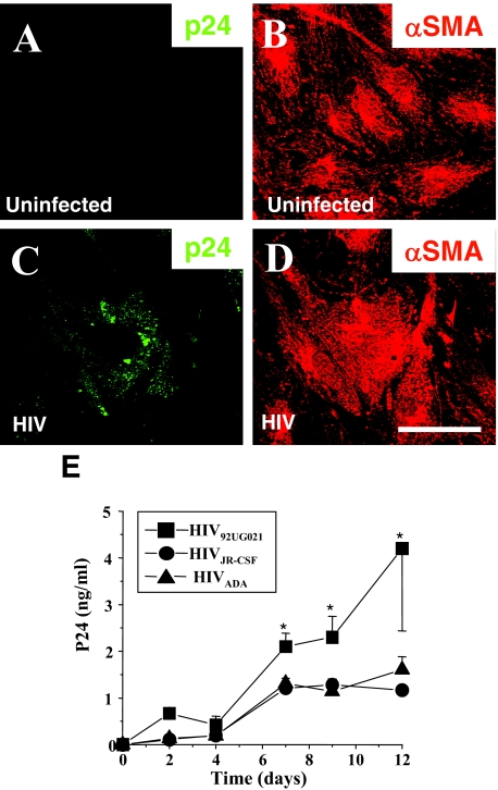
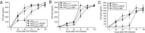
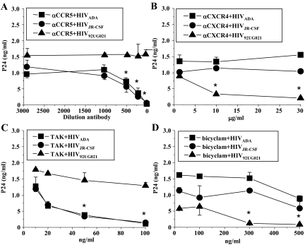
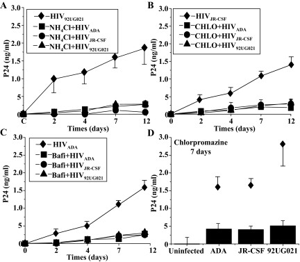
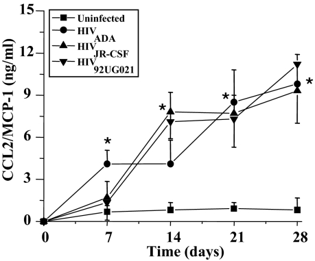
Human immunodeficiency virus (HIV) infection is associated with accelerated atherosclerosis and vasculopathy, although the mechanisms underlying these findings have not been determined. Hypotheses for these observations include: 1) an increase in the prevalence of established cardiac risk factors observed in HIV-infected individuals who are currently experiencing longer life expectancies; 2) the dyslipidemia reported with certain HIV anti-retroviral therapies; and/or 3) the proinflammatory effects of infiltrating HIV-infected monocytes/macrophages. An unexplored possibility is whether HIV itself can infect vascular smooth muscle cells (SMCs) and, by doing so, whether SMCs can accelerate vascular disease. Our studies demonstrate that human SMCs can be infected with HIV both in vivo and in vitro. The HIV protein p24 was detected by fluorescence confocal microscopy in SMCs from tissue sections of human atherosclerotic plaques obtained from HIV-infected individuals. Human SMCs could also be infected in vitro with HIV by a mechanism dependent on CD4, the chemokine receptors CXCR4 or CCR5, and endocytosis, resulting in a marked increase in SMC secretion of the chemokine CCL2/MCP-1, which has been previously shown to be a critical mediator of atherosclerosis. In addition, SMC proliferation appeared concentric to the vessel lumen, and minimal inflammation was detected, unlike typical atherosclerosis. Our data suggest that direct infection of human arterial SMCs by HIV represents a potential mechanism in a multifactorial paradigm to explain the exacerbated atherosclerosis and vasculopathy reported in individuals infected with HIV.


Figures

Figure 1
Figure 1
HIV infects SMCs in vivo. Immunohistochemical analyses of arteries from uninfected individuals without demonstrable plaque (denoted HIVneg/no plaque in A–D) or with atherosclerotic plaque (denoted HIVneg/plaque in E–H) were performed using antibodies to either human smooth muscle α-actin (SMA, red staining), HIV-p24 (green staining, p24), and DAPI (blue staining, nuclei), and were analyzed by confocal microscopy. DAPI staining was used to identify individual cells, blood vessel morphology, and cell accumulation (A, E, I, M). The arterial sections obtained from uninfected individuals without plaque (A–D) had a typical SMA staining (B) pattern with respect to the lumen (Lu) of the blood vessel and no detectable p24 staining (C). In sections obtained from uninfected individuals with plaque (E–H), SMC-rich areas (F) were present without detectable p24 antigen (G). Similar studies were done on atherosclerotic arterial sections from HIV-infected individuals (denoted HIVpos/plaque in I–P). In these samples, HIV p24 staining (K and O) was detected. The distribution of SMA and p24 appeared concentric with respect to the lumen (J) in the arterial sections with plaque from the HIV-infected individuals. At higher magnification, these sections (M–P) demonstrated the colocalization of the SMA-positive cells and p24 (P), as indicated by the arrows. A–L, scale bar = 25 μm; M–P, scale bar = 170 μm.
Figure 2
Figure 2
HIV-infected tissue sections of atherosclerotic lesions have fewer monocyte/macrophages that are distinct from SMCs. Immunohistochemical analyses of arterial sections from uninfected individuals without demonstrable plaque (denoted HIVneg/no plaque in A–E) or with atherosclerotic plaque (denoted HIVneg/plaque in F–J) were performed using antibodies to human smooth muscle α-actin (SMA, red staining), HIV-p24 (green staining, p24), CD68 (Cyan staining, monocyte/macrophages), and DAPI (blue staining, nuclei), and then were analyzed by confocal microscopy. DAPI staining was used to identify individual cells, blood vessel morphology, and cell accumulation (A, F, K, P). The arterial sections obtained from uninfected individuals without plaque (A–E) had a typical SMA staining (B) pattern with respect to the lumen (Lu) of the blood vessel and no detectable p24 staining (C) and few macrophages, CD68 staining (D). In sections obtained from uninfected individuals with atherosclerotic plaque (F–J), SMC-rich areas (G) were present without detectable p24 antigen (H) and many monocyte/macrophages (I) were detectable, indicating inflammation. In atherosclerotic arterial sections from HIV-infected individuals (denoted HIVpos/plaque in K–O and P–T), HIV p24 staining (N, S) was detected. The distribution of SMA and p24 appeared concentric with respect to the lumen (L, Q) in the arterial sections with plaque from the HIV-infected individuals. CD68 staining in HIVpos/plaque individuals was minimal (N, S) compared to sections obtained from uninfected individuals (D, I) with atherosclerotic plaque. At higher magnification, these sections (P–T) demonstrated the colocalization of the SMA-positive cells and p24 (P), as indicated by the arrows. Some CD68 staining also colocalized with p24 staining (R, S, T, arrowheads), but the SMA-positive population of cells was distinct from the CD68-positive cells, indicating that HIV-infected SMCs in vivo are distinct from the monocyte/macrophages. A–O, scale bar = 25 μm; P–T, scale bar = 170 μm.
Figure 3
Figure 3
HIV infects SMCs in vitro. Human SMCs were stained with antibodies to either human smooth muscle α-actin (αSMA) (B, D) or p24 antigen (p24) (A, C) following infection for 7 days with either HIV92UG021 (C, D) or uninfected (A, B). Control uninfected SMCs did not show detectable p24 staining (A). SMCs exposed to HIV (C) had intracellular vesicular p24 staining, notably with the X4 virus; scale bar = 30 μm. E: HIV-infected SMCs produce increasing amounts of p24. To determine the p24 production following HIV exposure, human SMCs were exposed to either an R5 (HIVADA or HIVJR-CSF) or an X4 (HIV92UG021) virus for 2, 4, 7, 9, and 12 days. Supernatants were analyzed for p24 viral antigen by ELISA. No p24 was detected in uninfected cultures (data not shown). No significant differences were detected between the two R5 viruses and the X4 virus was significantly different from R5 viruses after 7 days postinfection (*P < 0.003, n = 7 independent experiments, mean ± SD).
Figure 4
Figure 4
CD4 participation in HIV infection of SMCs in vitro. Human SMCs were exposed to either an X4 (HIV92UG021) or an R5 (HIVADA or HIVJR-CSF) virus in the presence of blocking antibodies to CD4 (bCD4, 2.5 μg/ml). Blocking antibodies to CD4 were added to the SMC cultures 15 minutes before viral exposure. After 2 hours of viral exposure, the cells were washed and the supernatants were analyzed for p24 viral antigen by ELISA at 24, 48, 72, 96, and 120 hours after treatment. Significant differences were detected at 24, 48, and 72 hours postinfection with HIV92UG021 strain (A), HIVJR-CSF (B), and HIVADA (C) with the exception that HIVJR-CSF was not significantly different, P < 0.32, n = 3, at 72 hours, P < 0.32, n = 3. Significance was P < 0.005, n = 3. and further differences were in p24 accumulation were not detected at 168 and 288 hours postinfection (data not shown). Nonspecific effects were not detectable by using the nonimmune isotype IgG1 (IgG) (results expressed as means ± SD, n = 3).
Figure 5
Figure 5
HIV infection of SMCs in vitro is mediated by chemokine receptors. Human SMCs were exposed to either an R5 (HIVADA or HIVJR-CSF) or an X4 (HIV92UG021) virus. Blocking agents or antibodies were added to the SMC cultures 15 minutes before viral exposure. After 2 hours of viral exposure, the cells were washed and the supernatants were analyzed for p24 viral antigen by ELISA at 7 and 12 days after treatment. No significant differences were detected at 7 and 14 days. Representative data from 7 days are presented. Neutralizing antibodies to either human CCR5 (A, αCCR5; at concentrations 1:100, 1:200; 1:500, 1:1000, and 1:3000 dilution) or to human CXCR4 (B, αCXCR4; at concentrations 1, 10, and 30 μg/ml) resulted in an abrogation of p24 protein accumulation in the supernatants of SMCs treated with the R5 and X4 viruses, respectively. An inhibitor of binding to CCR5 (C), TAK-779 (TAK), at concentrations of 10, 20, 50, and 100 ng/ml, or an inhibitor of CXCR4 (D), the bicyclam (JM-2987), at concentrations of 10, 100, 300, and 500 ng/ml, also reduced the p24 production in the supernatants by R5 or X4 viruses, respectively (results expressed as means ± SD).
Figure 6
Figure 6
HIV infection of SMCs in vitro is mediated by endocytosis and clathrin-mediated endocytosis. Human SMCs were exposed for 2 hours to either an R5 (HIVADA or HIVJR-CSF) or an X4 (HIV92UG021) virus in the presence of three general inhibitors of endocytosis, NH4Cl (50 mmol/L) (A), chloroquine (Chlo, 100 μg/ml) (B), bafilomycin-A (Bafi, 50 nmol/L) (C), and chlorpromazine, a clathrin-specific inhibitor of endocytosis (D). All inhibitors were added to SMCs 15 minutes before viral exposure. Viral replication was determined by assaying p24 antigen in the tissue culture media at 2, 4, 7, 9, and 12 days after HIV exposure. Inhibitors of endocytosis abolished the entry of all viruses tested (A–D) (n = 4 independent experiments). A representative experiment illustrating SMCs infected with HIV92UG021 (A), HIVJR-CSF (B), or HIVADA (C) in the absence of inhibitors (♦) is shown. These inhibitors had no effect on p24 levels in supernatants from human PBMCs that had been exposed to these isolates (data not shown) (results expressed as means ± SD).
Figure 7
Figure 7
HIV infection of SMCs increases CCL2/MCP-1 production. SMCs in vitro were exposed to one of the three strains HIVADA, HIVJR-CSF, or HIV92UG021 for 1 to 2 hours. Cells were washed extensively and then fresh medium placed. Media was collected and assayed by quantitative sandwich ELISA for CCL2 after 7, 14, 21, and 28 days postinfection. Significant amounts of CCL2 (*) were detected in the SMC supernatants of HIV-infected cultures at 14 days and from the ADA isolate beginning at 7 days postinfection. No differences in the amount of CCL2 production were noted among the three HIV isolates tested. A P value <0.05 is considered significant (*) as compared to untreated SMCs (n = 3 independent experiments, mean ± SD).

References

    1. Bokazhanova A, Rutherford GW. The epidemiology of HIV and AIDS in the world. Coll Antropol. 2006;30(Suppl 2):3–10. - PubMed
    1. de Saint Martin L, Vandhuick O, Guillo P, Bellein V, Bressollette L, Roudaut N, Amaral A, Pasquier E. Premature atherosclerosis in HIV positive patients and cumulated time of exposure to antiretroviral therapy (SHIVA study). Atherosclerosis. 2006;185:361–367. - PubMed
    1. Hsue PY, Giri K, Erickson S, MacGregor JS, Younes N, Shergill A, Waters DD. Clinical features of acute coronary syndromes in patients with human immunodeficiency virus infection. Circulation. 2004;109:316–319. - PubMed
    1. Segev A, Cantor WJ, Strauss BH. Outcome of percutaneous coronary intervention in HIV-infected patients. Catheter Cardiovasc Interv. 2006;68:879–881. - PubMed
    1. van Wijk JP, de Koning EJ, Cabezas MC, Joven J, op’t Roodt J, Rabelink TJ, Hoepelman AM. Functional and structural markers of atherosclerosis in human immunodeficiency virus-infected patients. J Am Coll Cardiol. 2006;47:1117–1123. - PubMed

Publication types

MeSH terms