. 2008 Apr;172(4):1030-42.
doi: 10.2353/ajpath.2008.070605. Epub 2008 Feb 29.

Blockade of endothelinergic receptors prevents development of proliferative vitreoretinopathy in mice

Affiliations

Blockade of endothelinergic receptors prevents development of proliferative vitreoretinopathy in mice

María Iribarne et al. Am J Pathol. 2008 Apr.

Abstract

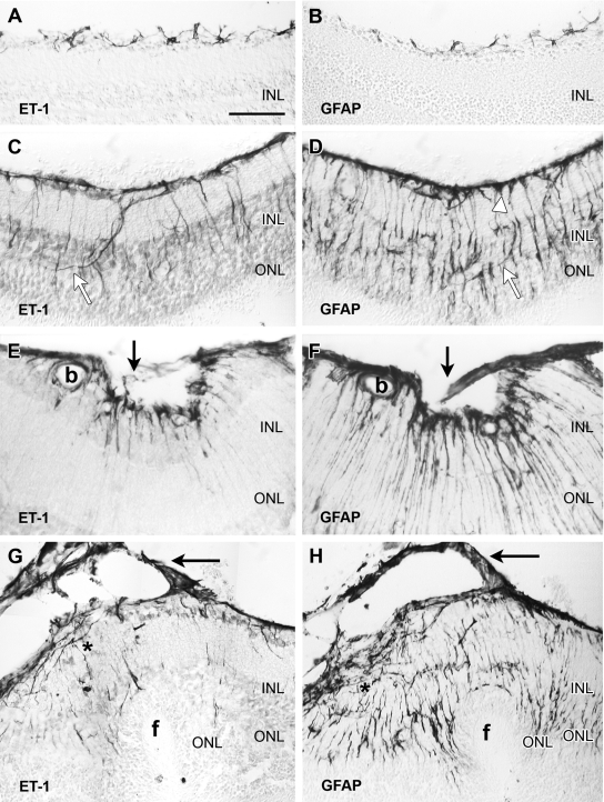
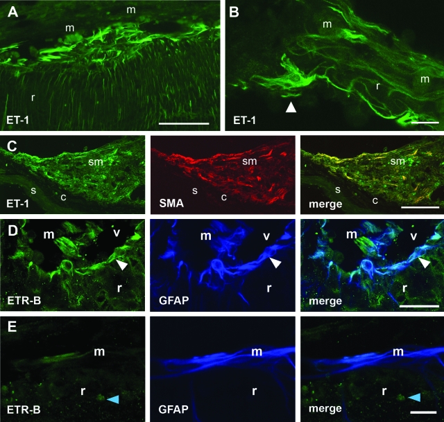
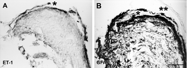
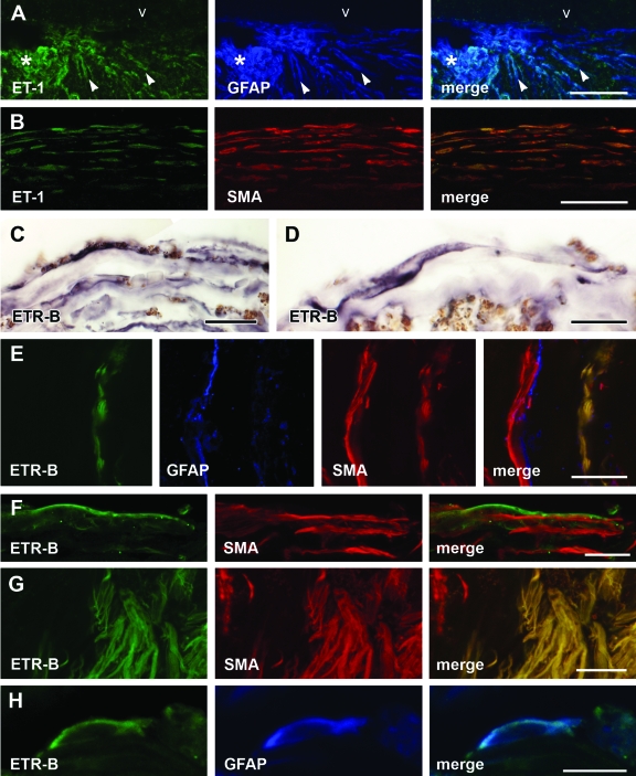
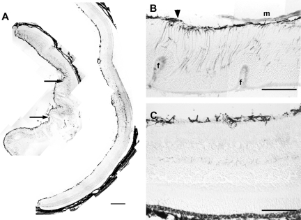
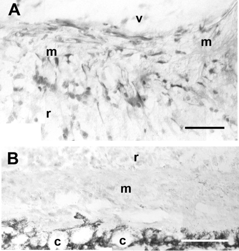
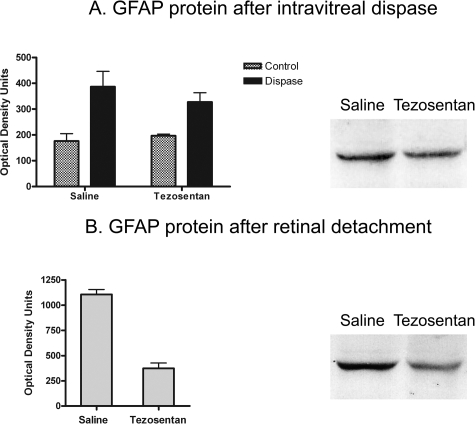
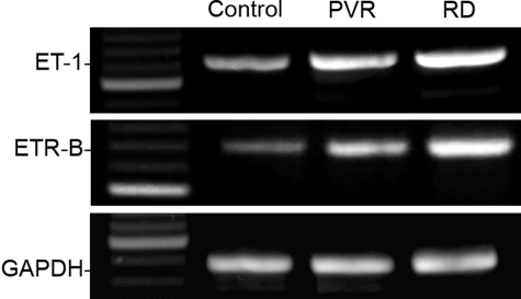
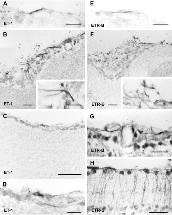
Proliferative vitreoretinopathy (PVR) is characterized by severe glial remodeling. Glial activation and proliferation that occur in brain diseases are modulated by endothelin-1 (ET-1) and its receptor B (ETR-B). Because retinal astrocytes contain ET-1 and express ETR-B, we studied the changes of these molecules in an experimental mouse model of PVR and in human PVR. Both ET-1 and ETR-B immunoreactivities increased in mouse retina after induction of PVR with dispase. Epi- and subretinal outgrowths also displayed these immunoreactivities in both human and experimental PVR. Additionally, myofibroblasts and other membranous cell types showed both ET-1 and ETR-B immunoreactivities. In early stages of experimentally induced PVR, prepro-ET-1 and ETR-B mRNA levels increased in the retina. These mRNA levels also increased after retinal detachment (RD) produced by subretinal injection. Treatment of mice with tezosentan, an antagonist of endothelinergic receptors, reduced the histopathological hallmarks of dispase-induced PVR: retinal folding, epiretinal outgrowth, and gliosis. Our findings in human and in dispase-induced PVR support the involvement of endothelinergic pathways in retinal glial activation and the phenotypic transformations that underlie the growth of membranes in this pathology. Elucidating these pathways further will help to develop pharmacological treatments to prevent PVR. In addition, the presence of ET-1 and ETR-B in human fibrous membranes suggests that similar treatments could be helpful after PVR has been established.


Figures

Figure 1
Figure 1
Consecutive sections of mouse retina immunoenzymatically stained with antibodies against ET-1 (left) and GFAP (right). The retinal vitreal surface is always shown at the top of the micrographs. A and B: In the normal retina, ET-1 immunoreactivity was only found in astrocytes sparsely distributed along the vitreal surface. GFAP immunostaining labeled similar cell bodies and processes. C and D: These and following images correspond to 0.2 U/μl dispase-injected eyes. One week after injection, the vitreal surface was completely covered by ET-1-immunoreactive astrocyte cell bodies and processes. Irregular ET-1-immunoreactive processes, following a nonradial course, extended into outer layers of the retina (white arrows). GFAP immunolabeled structures along the vitreal surface included astrocytes and Müller endfeet (white arrowhead). Notice the large number of GFAP-immunoreactive processes extending to the outer retinal layers. Most of them followed the typical radial course of Müller cells, but a few resembled endothelinergic processes (white arrows). E and F: Illustration of an inner retinal fold bridged by a small outgrowth (arrows), appearing a week after dispase injection. Strong ET-1 and GFAP immunoreactivities appeared along the vitreal border, within the outgrowth and in retinal processes. GFAP revealed a larger number of processes than ET-1. Immunostained glial cells also surrounded a blood vessel (b). G and H: Retina sections (2 weeks after dispase) showing an outer fold (f). A thin cellular membrane (arrows) attached to the vitreal surface exhibited strong ET-1 and GFAP immunostaining. Elongate ET-1- and GFAP-immunoreactive cells were accumulated along the retinal vitreal surface (asterisk). Immunoreactive processes extended across the retina: few endothelinergic processes reached the invaginated ONL, whereas numerous GFAP-immunoreactive processes appeared close to the subretinal folded surface. Scale bar: 50 μm (A–E, G, H); 25 μm (F).
Figure 2
Figure 2
ET-1 and ETR-B immunofluorescence in epi- and subretinal outgrowths appearing 2 weeks after 0.2 U/μl dispase intravitreal injection. A: Strong ET-1 immunofluorescence appeared in a region of disrupted ILM, where the vitreal surface was attached to an epiretinal membrane (m). Notice the thick endothelinergic processes parallel to the surface and the large number of thinner processes within the retina. B: In a membrane attached to the vitreal surface, an ET-1-immunoreactive cell could be identified as an astrocyte by its shape and planar distribution of cell processes. Numerous endothelinergic processes appeared within the membrane. r, retina. C: Confocal images of a subretinal membrane (sm) attached to the choroid (c). Numerous cells exhibiting ET-1 (green) and SMA (red) immunofluorescence can be observed. Merging demonstrates co-localization of both markers in elongate cells probably corresponding to myofibroblasts. s, sclera. D: Confocal images through the inner retina (r) showed ETR-B-immunoreactive cell bodies and processes placed along the vitreal surface (white arrowhead). GFAP antiserum labeled the same structures. Some radial processes were labeled by only one of these markers. An epiretinal membrane (m) appearing within the vitreal (v) space showed almost complete segregation of ETR-B and GFAP immunofluorescence. E: Confocal images through an epiretinal membrane attached to the retinal surface showed a few ETR-B-immunoreactive cells. GFAP labeled a larger group of elongate cells. The merged image demonstrated that these ETR-B-immunoreactive cells also had GFAP immunoreactivity, whereas most GFAP-immunoreactive cells lacked ETR-B immunofluorescence. The blue arrowhead points to an ETR-B-immunoreactive cell nucleus. Scale bars: 50 μm (A, B); 100 μm (C); 25 μm (D, E).
Figure 3
Figure 3
Sections through a retinal fragment embedded in a human PVR specimen labeled with the immunoenzymatic method and examined under Nomarski optics. A: ET-1 immunoreactivity appeared along the vitreal surface of a highly disorganized retinal fragment. An epiretinal outgrowth (asterisk) separated from the inner retinal surface also showed strong ET-1 immunoreactivity. B: A section through the same fragment (not consecutive) showed extensive GFAP immunoreactivity in most retinal layers. GFAP-immunoreactive cells were present in an outgrowth (asterisk) apparently emerging from the retinal surface. A neighboring outgrowth (asterisks) had no immunoreactivity. Scale bar: 50 μm.
Figure 4
Figure 4
ET-1- and ETR-B-immunoreactive cells in human specimens. A: Confocal images through a retinal fragment embedded in a human epiretinal membrane showed an epiretinal outgrowth (asterisk) displaying strong ET-1 immunoreactivity. ET-1-immunoreactive cells followed radial pathways across the retina (arrowheads) or coursed along its vitreal surface (v). GFAP immunofluorescence essentially labeled the same structures. However, some GFAP-immunoreactive cells lacked ET-1 immunofluorescence. B: Confocal images of a fibrous human epiretinal membrane showing elongate cells arrayed in parallel layers. These cells displayed both ET-1 and SMA immunofluorescence as demonstrated in the merged image. C: The immunoenzymatic procedure demonstrated elongate ETR-B-labeled arrayed in parallel layers. D: This view of a fibrous membrane showed a long ETR-B-immunoreactive cell parallel to the surface of the membrane. The membrane contained abundant pigmented granules. E: Confocal images of a fibrous membrane showed a layer of ETR-B cells. A different cell layer displayed GFAP immunofluorescence. Both layers had SMA immunoreactivity. ETR-B co-localized with SMA in cells of the first layer (right). In the second layer, GFAP immunofluorescence was segregated from SMA. F: Elongate cells displayed ETR-B immunofluorescence. SMA immunoreactivity appeared in cells of similar shape. However, no co-localization could be detected in these cells. G: Another membrane showed a large cluster of ETR-B-immunoreactive cells. The same cells also displayed SMA immunofluorescence. However, a neighboring group of SMA-immunoreactive cells (left) was not labeled by ETR-B. H: An isolated cell displaying ETR-B immunofluorescence also showed GFAP immunoreactivity. Merge demonstrates co-localization. Scale bars: 25 μm (A, C, E–G); 50 μm (B); 15 μm (D, H).
Figure 5
Figure 5
A: These low-magnification montages of GFAP-immunostained retinal cryosections show the effect of tezosentan treatment on retinal lesions appearing 1 week after 0.3 U/μl dispase injection. The retina from an untreated mouse (left) displayed severe folding. Arrows point to epiretinal membranes. Large areas of the inner retina displayed strong GFAP immunoreactivity. The retina from a mouse receiving tezosentan (right) had no folds. The vitreal surface showed increased GFAP immunoreactivity. GFAP immunostaining of the inner retina was only found at a small region of the retina. B: Outer retinal folds (f) and an epiretinal membrane were present in this untreated retina. GFAP-immunoreactive structures form a thick layer along the vitreal surface and extend toward the outer retina. The arrowhead points to a GFAP-immunoreactive vitreal outgrowth. C: This retina from a treated animal appeared well attached to the RPE and showed good preservation of retinal layering. GFAP-immunoreactive structures were restricted to the vitreal surface. Scale bars: 200 μm (A); 60 μm (B, C).
Figure 6
Figure 6
ETR-B immunoreactivity in mouse epi- and subretinal membranes developing 1 week after 0.3 U/μl dispase injection. A: This epiretinal membrane (m) was attached to the retinal surface and exhibited numerous ETR-B-immunoreactive cells. v, vitreal space; r, retina. B: A subretinal membrane (m), lodged between the retina (r) and the choroid (c), lacked ETR-B immunostaining. Scale bars: 50 μm (A); 100 μm (B).
Figure 7
Figure 7
A: Differences of GFAP protein levels in control and dispase-injected eyes, receiving saline or tezosentan (each group, n = 4), were evaluated with two-way analysis of variance. Tezosentan did not increase GFAP protein in control mice. Dispase injection (0.3 U/μl) produced a significant increase of GFAP protein (P < 0.01). This increase was not affected by tezosentan treatment. Bands corresponding to a pair of saline- and tezosentan-treated dispase-injected eyes showed similar immunostaining. B: A large increase of GFAP protein followed RD. Tezosentan treatment significantly reduced these levels. Paired t-test (n = 8 pairs, P < 0.0004). The bands corresponding to a pair of saline- and tezosentan-treated RD samples illustrate this large reduction.
Figure 8
Figure 8
Effect of experimental PVR and RD on amplification of retinal prepro-ET-1, ETR-B, and GADPDH mRNA sequences. Extracts were prepared 3 days after each procedure. Intensity of the prepro-ET-1 and ETR-B amplification products increased after experimental PVR and RD. Both bands were always stronger after RD than after PVR. Intensity of the GAPDH amplification product did not change significantly.
Figure 9
Figure 9
Immunoreactivity of ET-1 (left) and ETR-B (right) was demonstrated by the immunoenzymatic procedure 3 days after RD or dispase injection (0.3 U/μl). Images were acquired with Nomarski optics. A: This high-power view, corresponding to a normal retina, showed that isolated astrocytic processes were present. B: A retinal fold induced by dispase injection, showed numerous ET-1-immunostained astrocytes. The ILM had disappeared and astrocyte processes invaded the vitreal space. The inset illustrates the large size of these astrocytes. C and D: Low- and high-power views of ET-1 immunoreactivity after RD. ET-1 was moderately increased and could only be detected in astrocytes. E: This high-power view showed weak ETR-B immunoreactivity in astrocytic processes of control retinas. F: After dispase injection, numerous ETR-B-immunoreactive astrocytes appeared along the vitreal surface of the retina. In regions of ILM disruption, numerous astrocytic processes extended into the vitreal space. G and H: These images correspond to detached retinas. Notice the preservation of the ILM in both examples. G shows perivascular astrocytes displaying strong ETR-B immunoreactivity. H shows a retinal region displaying ETR-B immunoreactivity in astrocytes and Müller cells. Cell nuclei were also strongly immunostained in these preparations. Scale bars: 15 μm [A, B (inset), D, E, F (inset)]; 25 μm (B, F–H); 50 μm (C).

Similar articles

Cited by

References

    1. Charteris DG, Sethi CS, Lewis GP, Fisher SK. Proliferative vitreoretinopathy—developments in adjunctive treatment and retinal pathology. Eye. 2002;16:369–374. - PubMed
    1. Scott IU, Flynn HW, Jr, Murray TG, Feuer WJ. Outcomes of surgery for retinal detachment associated with proliferative vitreoretinopathy using perfluoro-n-octane: a multicenter study. Am J Ophthalmol. 2003;136:454–463. - PubMed
    1. Fisher SK, Lewis GP, Linberg KA, Verardo MR. Cellular remodeling in mammalian retina: results from studies of experimental retinal detachment. Prog Retin Eye Res. 2005;24:395–431. - PubMed
    1. Sethi CS, Lewis GP, Fisher SK, Leitner WP, Mann DL, Luthert PJ, Charteris DG. Glial remodeling and neural plasticity in human retinal detachment with proliferative vitreoretinopathy. Invest Ophthalmol Vis Sci. 2005;46:329–342. - PubMed
    1. Lewis GP, Fisher SK. Muller cell outgrowth after retinal detachment: association with cone photoreceptors. Invest Ophthalmol Vis Sci. 2000;41:1542–1545. - PubMed

Publication types

MeSH terms