. 2008 Apr;5(4):331-8.
doi: 10.1038/nmeth.1187. Epub 2008 Mar 2.

Photochemical control of endogenous ion channels and cellular excitability

Affiliations

Photochemical control of endogenous ion channels and cellular excitability

Doris L Fortin et al. Nat Methods. 2008 Apr.

Abstract

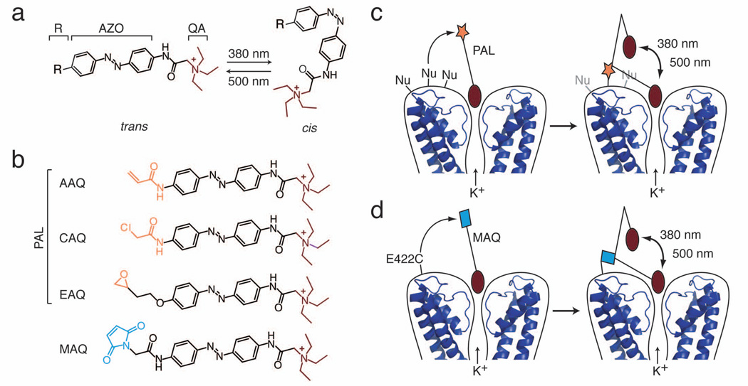
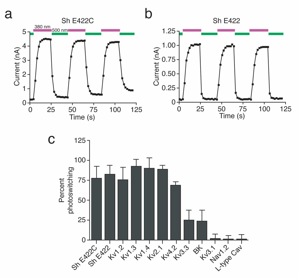
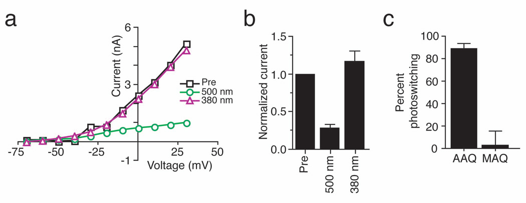
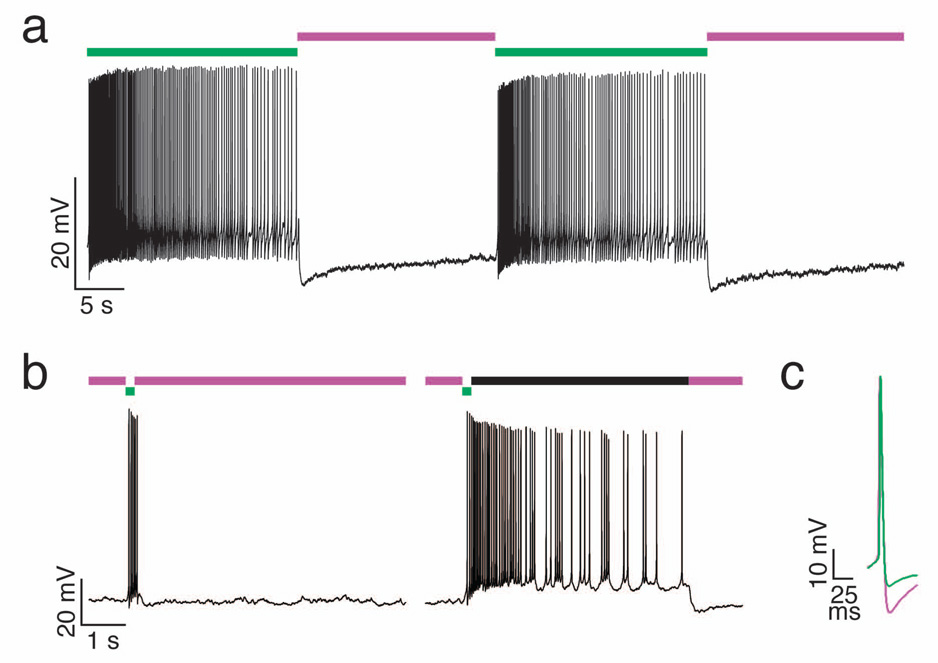
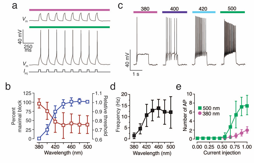
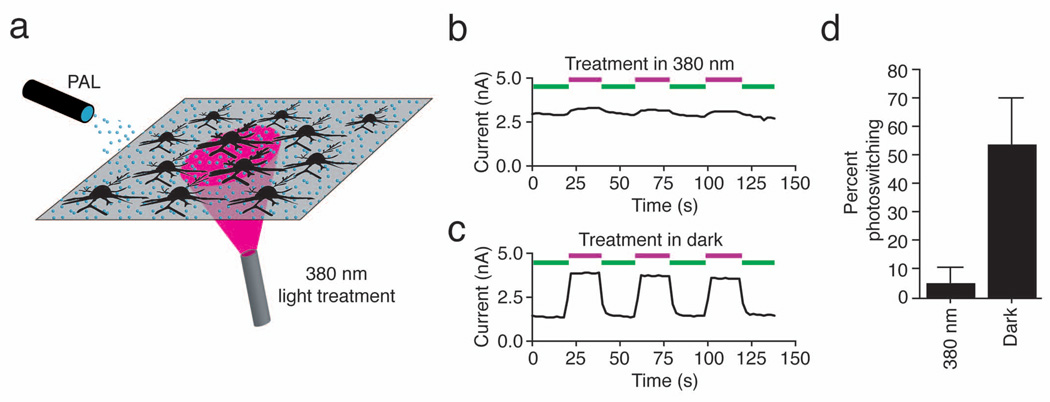
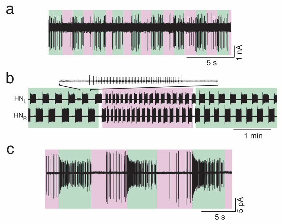
Light-activated ion channels provide a precise and noninvasive optical means for controlling action potential firing, but the genes encoding these channels must first be delivered and expressed in target cells. Here we describe a method for bestowing light sensitivity onto endogenous ion channels that does not rely on exogenous gene expression. The method uses a synthetic photoisomerizable small molecule, or photoswitchable affinity label (PAL), that specifically targets K+ channels. PALs contain a reactive electrophile, enabling covalent attachment of the photoswitch to naturally occurring nucleophiles in K+ channels. Ion flow through PAL-modified channels is turned on or off by photoisomerizing PAL with different wavelengths of light. We showed that PAL treatment confers light sensitivity onto endogenous K+ channels in isolated rat neurons and in intact neural structures from rat and leech, allowing rapid optical regulation of excitability without genetic modification.


Figures

Figure 1
Figure 1. The PAL approach for imparting light sensitivity onto native ion channels
(a) PAL molecules consist of a photoisomerizable azobenzene group (AZO) flanked by a quaternary ammonium (QA; burgundy) and a covalent attachment group (R). Exposure to 380 nm light isomerizes the AZO to its shorter cis form whereas exposure to 500 nm light favors the trans configuration. (b) PAL molecules contain a promiscuous reactive group (orange): acrylamide (Acryl-AZO-QA or AAQ), chloroacetamide (CAQ), or epoxide (EAQ). In contrast, MAQ contains a maleimide (blue) designed to react with an engineered cysteine. (c) After QA binds to the channel pore, PAL reacts via its promiscuous reactive group with an endogenous nucleophile (Nu) on native K+ channels. Once tethered, the photoswitch allows control of ionic current using light. In 500 nm light, the photoswitch is extended, blocking ion conduction. In 380 nm light, AZO isomerizes to its cis form, retracting the QA and allowing ion conduction. Molecular coordinates from KcsA (PDB ID: 2A9H) were drawn using MacPyMol (DeLano, W.L. The PyMOL Molecular Graphics System (2002); http://www.pymol.org). (d) To generate the SPARK channel, the maleimide group of MAQ attaches to a cysteine (E422C) genetically engineered in a Shaker K+ channel.
Figure 2
Figure 2. Photocontrol of K+ channels expressed in HEK293 cells
(a) AAQ photosensitizes Shaker channels that contain an engineered nucleophilic attachment site (Sh E422C). Voltage-gated K+ currents were elicited by pulsing from −70 to +30 mV for 250 ms. 500 nm light (green) blocks current through the channels whereas 380 nm light (violet) unblocks the channels. (b) AAQ photosensitizes wild-type Shaker channels (Sh E422). Pulse protocols as in (a). (c) Percent photoswitching for different channels treated with AAQ (400 µM, 15 min). We defined percent photoswitching as the difference between the steady-state current in 380 and 500 nm light, divided by the current in 380 nm light. The extent of photoswitching is similar for Shaker with or without the engineered cysteine (n = 4 for each). AAQ treatment also strongly photosensitizes Kv1.2, 1.3, 1.4, 2.1, and 4.2. Currents through Kv3.3 and BK are only modestly affected by light after AAQ treatment. Kv3.1 is insensitive to AAQ as are voltage-gated Na+ channels expressed in HEK293T cells and L-type Ca2+ channels endogenous to GH3 cells (n = 4–7 cells for each channel type). Current were elicited by stepping from −70 to +30 mV (K+ channels), −80 to 0 mV (Na+ channels), and −40 to +20 mV (Ca2+ channels).
Figure 3
Figure 3. Photocontrol of native K+ current in cultured hippocampal neurons
(a) Steady-state I–V curves from a voltage-clamped hippocampal pyramidal neuron before (Pre; black squares) and after a 15 min application of 300 µM AAQ. Photoisomerization of AAQ to the trans state with 500 nm light (green circles) reduces voltage-gated K+ current whereas illumination with 380 nm light (violet triangles) restores current to levels similar to those measured prior to AAQ treatment. (b) AAQ-treated channels are completely unblocked by 380 nm light. Voltage-gated currents (elicited by stepping from −70mV to +30mV) measured after AAQ treatment were normalized to those measured prior to AAQ application (n = 5). (c) Percent photoswitching for K+ current in hippocampal neurons treated with AAQ (200 µM) or MAQ (250 µM) (n = 6 for each). Light has no effect on the steady-state I–V curve from a non-transfected neuron treated with MAQ. MAQ only imparts light-sensitivity on neurons transfected with the Shaker E422C channel.
Figure 4
Figure 4. Photocontrol of neuronal firing
(a) Current clamp recording from a hippocampal pyramidal neuron treated with AAQ. Depolarizing current (90 pA) was injected to induce continuous action potential firing in 500 nm light (green). Illumination with 380 nm light (violet) rapidly suppresses action potential firing. High frequency firing resumes upon illumination with 500 nm light. (b) Current clamp recording from a hippocampal pyramidal neuron treated with AAQ. Depolarizing current (15 pA) was injected to bring the cell close to threshold. Left: a 200 ms flash of 500 nm light (green) induces a transient burst of action potential that is terminated rapidly upon illumination with 380 nm light (violet). Right: a persistent burst of action potential that outlast the light stimulus can be generated when the cell is illuminated briefly with 500 nm light (green) followed by darkness (black bar). Firing ceases once the neuron is illuminated with 380 nm light (violet). (c) Block of K+ channels with AAQ in 500 nm light (green) decreases the action potential afterhyperpolarization compared to that under 380 nm light (violet). Action potentials were induced with 500 ms depolarizing current injection (120 pA in 380 nm light, 100 pA in 500 nm light).
Figure 5
Figure 5. Modulation of neuronal excitability with light
(a) PAL imparts photocontrol over the current threshold for eliciting action potentials in hippocampal neurons. Current pulses (250 pA, 50 ms; 5 Hz) that fail to induce spikes in 380 nm light, reliably elicit spikes in 500 nm light. (b) Control of voltage-gated K+ channel blockade and action potential threshold by different wavelengths of light. Graded block of voltage-gated K+ current with increasing wavelengths of light (blue, n = 5 cells) decreases the current injection threshold for eliciting spikes (red, n = 6 cells). (c) PAL imparts photocontrol over spike frequency adaptation. Current injection (300 pA, 1s) triggers an adapting response consisting of a single spike when K+ channels are unblocked with 380 nm light. K+ channel blockade with longer wavelength light (indicated above colored bars) gradually decreases spike frequency adaptation, allowing the same current injection to elicit repetitive, high frequency firing. (d) Relationship between illumination wavelength and average spike frequency during depolarizing pulses (n = 9 cells). (e) Blockade of PAL-modified channels using 500 nm light decreases firing threshold and increases the number of action potentials fired in response to a given current injection (n = 6 cells. Currents were normalized to the maximal amount of current injected in each cell. Current injections lasted 500 ms and ranged from 15 to 360 pA).
Figure 6
Figure 6. Local illumination during PAL treatment imprints photosensitivity onto specific neurons
(a) Illustration of experimental setup. A hippocampal neuronal culture was uniformly exposed to 300 µM AAQ. The microscope objective was used to illuminate a subpopulation of neurons with 380 nm light with the remaining neurons were kept in darkness. After treatment, AAQ was replaced with extracellular solution and whole-cell recordings were obtained. (b) Photosensitization of voltage-gated outward current is prevented in a neuron locally exposed to 380 nm light during AAQ treatment. Currents were elicited by stepping from −70 mV to +30 mV for 250 ms every 2 sec. (c) Photosensitization of voltage-gated outward current is robust in a neuron kept in darkness during AAQ treatment. Recordings in panels b and c were from neurons on the same coverslip. (d) Exposure to 380 nm light during AAQ treatment prevents photosensitization of K+ channels (n = 9 and 10 neurons for reaction in the dark and under 380 nm light respectively).
Figure 7
Figure 7. Photocontrol of action potential firing in intact circuits
(a) Recording from a cerebellar basket cell. Unblocking AAQ-modified K+ channels with 360 nm light (violet) prevents basket cell firing while blocking AAQ-modified K+ channels with 500 nm light (green) depolarizes basket cells, resulting in action potential firing. (b) Simultaneous loose-patch recordings of bursting activity from the left and right heart interneurons (HNL and HNR) in the leech heart beat CPG. Blocking AAQ-modified K+ channels with 500 nm light (green) increased the bursting cycle period. Unblocking AAQ-modified channels with 380 nm light (violet) decreased the period. (c) Loose-patch recording of PAL-mediated photoresponses in a rat RGC from a flat-mounted retina. Blocking AAQ-modified K+ channels with 500 nm light (green) promotes high-frequency firing; 380 nm light (violet) inhibits firing.

Comment in

References

    1. Callaway EM, Yuste R. Stimulating neurons with light. Curr. Opin. Neurobiol. 2002;12:587–592. - PubMed
    1. Kramer RH, Chambers JJ, Trauner D. Photochemical tools for remote control of ion channels in excitable cells. Nat. Chem. Biol. 2005;1:360–365. - PubMed
    1. Miesenbock G, Kevrekidis IG. Optical imaging and control of genetically designated neurons in functioning circuits. Annu. Rev. Neurosci. 2005;28:533–563. - PubMed
    1. Zhang F, Wang LP, Boyden ES, Deisseroth K. Channelrhodopsin-2 and optical control of excitable cells. Nat. Methods. 2006;3:785–792. - PubMed
    1. Ellis-Davies GC. Caged compounds: photorelease technology for control of cellular chemistry and physiology. Nat. Methods. 2007;4:619–628. - PMC - PubMed

Publication types

MeSH terms