. 2008 Mar;15(3):303-11.
doi: 10.1038/nsmb.1394. Epub 2008 Mar 2.

GroEL stimulates protein folding through forced unfolding

Affiliations

GroEL stimulates protein folding through forced unfolding

Zong Lin et al. Nat Struct Mol Biol. 2008 Mar.

Abstract

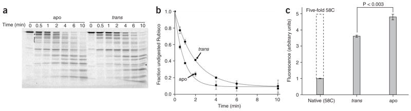
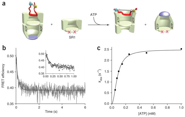
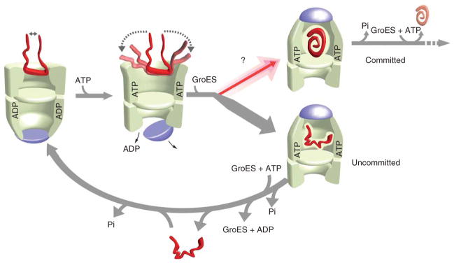
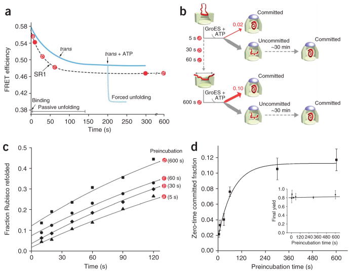
Many proteins cannot fold without the assistance of chaperonin machines like GroEL and GroES. The nature of this assistance, however, remains poorly understood. Here we demonstrate that unfolding of a substrate protein by GroEL enhances protein folding. We first show that capture of a protein on the open ring of a GroEL-ADP-GroES complex, GroEL's physiological acceptor state for non-native proteins in vivo, leaves the substrate protein in an unexpectedly compact state. Subsequent binding of ATP to the same GroEL ring causes rapid, forced unfolding of the substrate protein. Notably, the fraction of the substrate protein that commits to the native state following GroES binding and protein release into the GroEL-GroES cavity is proportional to the extent of substrate-protein unfolding. Forced protein unfolding is thus a central component of the multilayered stimulatory mechanism used by GroEL to drive protein folding.


Figures

Figure 1
Figure 1
Non-native Rubisco is more compact when bound to the trans ring of an ADP bullet than when bound to apoGroEL. (a) The conformation of a substrate protein bound to either apoGroEL or the trans ring of an ADP bullet can be investigated with intramolecular FRET, where the distance between two exogenous fluorescent probes attached to different parts of the substrate protein (indicated schematically as colored circles labeled ‘A’ and ‘D’) is examined. (b) Steady-state FRET measurement of the distance between the N- and C-terminal domains of a non-native Rubisco monomer (100 nM) bound to an ADP-bullet trans ring and apoGroEL (120 nM in each case). The C-terminal domain of a previously described Rubisco variant was labeled with a donor fluorophore and the N-terminal domain was labeled with an acceptor fluorophore. For these measurements, the donor probe was 5-(2-acetamidoethyl) aminonaphthalene-1-sulfonate (AEDANS; 454-ED) and the acceptor was fluorescein (58-F). The donor-emission spectra of donor-only (labeled apo-D and trans-D, respectively) and donor-acceptor (labeled apo and trans, respectively) samples are shown. (c) Time-resolved donor-intensity decays of the samples described in b. The donor-emission decay of donor-only and donor-acceptor samples, labeled as above, and the instrument response function (IRF) are shown. The average intraprobe distance was quantified from both steady-state and time-resolved FRET measurements (b, inset).
Figure 2
Figure 2
Non-native Rubisco bound to the trans ring of an ADP bullet is less susceptible to protease digestion and shows reduced chemical modification of internal cysteine residues. (a) Fluorescently labeled Rubisco (58-F; 100 nM) was denatured and bound to either ADP bullets or apoGroEL (labeled trans and apo, respectively; 120 nM in each case), treated with chymotrypsin for the indicated times and then analyzed by SDS-PAGE and laser-excited, fluorescence gel scanning. Partial digestion fragments that appear uniquely well represented in either the apo or trans digestion experiments are indicated by a square bracket and asterisks. (b) The amount of full-length, fluorescent Rubisco was quantified for three independent protease experiments, and the average of these replicates is plotted as a function of digestion time (error bars show one s.d.). The t1/2 for the disappearance of full-length Rubisco bound to apoGroEL and an ADP-bullet trans ring are 0.6 min and 1.3 min, respectively. (c) The internal structure of Rubisco monomers bound to apoGroEL and the ADP-bullet trans ring was probed with a small, highly reactive coumarin maleimide dye (CPM) that selectively modifies exposed cysteine residues. Only a single surface-exposed cysteine residue at position 58 (out of a total of five cysteine residues in wild-type Rubisco) is modified in native Rubisco (labeled Native (58C); 500 nM). The graph shows the total level of CPM fluorescence detected in the Rubisco sample following analysis by reverse-phase HPLC. When non-native Rubisco (500 nM) is bound to either apoGroEL (apo; 600 nM) or an ADP-bullet trans ring (trans; 600 nM), the internal Rubisco cysteine residues become highly reactive toward CPM. However, the reactivity of internal cysteine residues in Rubisco bound to apoGroEL is significantly greater than the trans ring–bound sample (P < 0.003 based on a paired t-test with n = 3 replicates; error bars show one s.d.). The maximum level of internal CPM incorporation expected under the solution conditions used is also shown (indicated by a dashed line, labeled five-fold 58C) and was estimated by assuming that the internal Rubisco cysteine residues in unfolded Rubisco would be as exposed and reactive as the surface-exposed 58C site.
Figure 3
Figure 3
GroES and ATP binding to a Rubisco-occupied trans ring results in forced conformational expansion, followed by compaction, of the non-native protein. (a) Schematic of a FRET experiment designed to examine conformational changes in a non-native substrate protein upon encapsulation beneath GroES on the trans ring of an ADP bullet. (b) GroES binding and substrate encapsulation results in an initial rapid expansion of the non-native Rubisco intermediate, followed by a compaction. Substrate-bound complexes in the presence of excess GroES were rapidly mixed in a stopped-flow apparatus (60 nM) with ATP (1 mM). For these measurements, the donor probe was fluorescein and the acceptor was rhodamine. Each FRET trace is the average of n = 10 replicates of matched experimental pairs, calculated from donor-only (Rubisco 454-F) and donor-acceptor (Rubisco 454-F/58-R) samples. The inset shows the FRET change over the first second of data. The rising phase in the observed FRET signal reflects GroES binding and substrate-protein encapsulation and compaction. (c) The observed rate constants for the initial rapid drop in FRET efficiency (kobs) and the subsequent rise upon GroES binding (kES) for a series of experiments at different concentrations of GroES are shown. In each case, a fixed concentration of the Rubisco-bound GroEL ADP bullets (60 nM) and ATP (1 mM) was used, whereas the total concentration of GroES was varied.
Figure 4
Figure 4
ATP binding alone to a Rubisco-occupied trans ring drives a forced conformational expansion of the non-native protein. (a) Schematic of a FRET experiment designed to follow conformational changes in a non-native substrate protein upon ATP binding alone to the substrate-loaded trans ring of an ADP bullet. In this case, a large excess of a single-ringed GroEL variant SR1 (ref. 21) was added as a GroES trap. SR1 contains a set of equatorial mutations that prevent assembly of a double-ringed complex (indicated by red crosses). SR1 binds ATP and GroES, executes a single round of ATP hydrolysis and then stalls, failing to release GroES and ADP. (b) The allosteric transition triggered by ATP alone results in the forced conformational expansion of the non-native protein. Stopped-flow FRET experiments were conducted in essentially the same manner as in Figure 3, except that SR1 was present in a 20-fold excess (1.2 μM) to prevent GroES binding to the substrate-loaded GroEL ring. (c) The observed rate of Rubisco expansion on the trans ring of an ADP bullet at different concentrations of ATP is shown. Each data point in the plot represents a single exponential fit to the observed change in FRET efficiency of the labeled Rubisco calculated from the average of n = 10 experimental replicates. The solid line shows a fit of the data to the Hill equation with kobs = 2.49 ± 0.06 s−1, K1/2 = 82.6 ± 3.7 μM and nH = 1.9 ± 0.2.
Figure 5
Figure 5
A functional model for substrate-protein unfolding by GroEL. Partial substrate unfolding by GroEL occurs in two phases: (i) passive unfolding upon protein capture by a GroEL ring; and (ii) forced unfolding upon ATP binding to a substrate-loaded trans ring. Although substrate confinement within the GroEL–GroES cavity enhances folding, the GroEL–GroES cavity exists for only a short period of time (~15–20 s at 25 °C) during a functional GroEL cycle,,,,,–. Substrate unfolding provides, in principle, a method for driving a non-native substrate protein to a higher level of its folding free energy landscape, opening efficient folding paths (committed) that are not readily accessible to more compact states (uncommitted),. Substrate-protein unfolding before encapsulation might substantially enrich the fraction of protein that commits to a productive folding path with each round of the GroEL cycle. ATP binding to the trans ring of the asymmetric complex, once hydrolysis of ATP within the GroEL–GroES cavity is complete (Pi), induces disassembly of the GroEL–ADP–GroES complex and ejection of the substrate protein, folded or not,,,. Uncommitted substrate must be recaptured on the trans ring of an ADP bullet for another round of unfolding and encapsulation (lower arrow). Two full rounds of ATP hydrolysis, one in each ring, followed by a round of substrate capture on the trans ring, returns the asymmetric ATP-containing complex to the starting ADP-bullet state.
Figure 6
Figure 6
ATP binding to a Rubisco-loaded SR1 ring does not cause extensive forced unfolding. (a) SR1 and the trans ring of an ADP bullet drive passive unfolding of Rubisco at a similar rate. The passive unfolding of Rubisco by SR1 and the ADP-bullet trans ring was monitored by FRET using fluorescein as the donor and rhodamine as the acceptor. The time-dependent drops in FRET efficiency for each complex are shown. Matched samples of donor-only (454-F) and donor-acceptor (454-F/58-R) Rubisco were denatured in acid-urea and rapidly diluted 50-fold (100 nM final) into buffer containing either SR1 (400 nM final) or ADP bullets (120 nM final) at 25 °C. Under the conditions used for this experiment, binding is complete within 5 s. In both cases, mixing was conducted by rapid manual mixing into a thermally jacketed stirring cuvette in a standard fluorimeter to allow stable long-time observation of the sample. (b) SR1 does not support extensive forced unfolding of non-native Rubisco. Acid-urea denatured Rubisco was diluted to a final concentration of 100 nM into buffer containing 400 nM SR1. At different times over the course of a passive unfolding curve (main plot), aliquots of the SR1–Rubisco complex were rapidly mixed with ATP in a stopped-flow apparatus, and changes in the FRET efficiency of the labeled Rubisco sample were monitored for ~10 s. The insets show representative stopped-flow traces (red) taken at early (left) and late (right) points in the passive-unfolding process. For reference, the progress of passive unfolding over the same time window is also shown in each inset. The stopped-flow FRET traces are the average of n = 3 replicates of matched experimental pairs, calculated from donor-only (Rubisco 454-F) and donor-acceptor (Rubisco 454-F/58-R) samples. (c) The maximum amplitude of forced Rubisco expansion by SR1 upon ATP binding was measured for a series of stopped-flow experiments over the full passive-unfolding curve and is plotted as a function of time. The level of preexisting passive unfolding does not affect the weak forced unfolding supported by SR1 upon ATP binding.
Figure 7
Figure 7
The fraction of non-native Rubisco that commits to the native state in a single round of GroES encapsulation increases in proportion to the extent of unfolding. (a) The slow, passive unfolding of Rubisco on SR1 can be used to examine the linkage between productive folding and substrate-protein unfolding. The progress curve of passive Rubisco unfolding by SR1, monitored by FRET at 25 °C, is shown schematically. For comparison, a schematic of the progress curve of Rubisco unfolding on the trans ring of an ADP bullet, and the extent of forced unfolding driven by ATP binding to the trans ring, are also shown. (b) Schematic of the experiment. Denatured Rubisco bound to excess SR1 was incubated for different preincubation times at 25 °C (indicated by red circles and letters a–d) to allow passive unfolding before the addition of GroES and ATP. Because SR1 shows a dramatically reduced capacity to support forced unfolding upon ATP binding (Fig. 6), the preincubation time primarily sets the level of unfolding imposed on the bound Rubisco monomer. Following addition of ATP and GroES, the encapsulated Rubisco is allowed to fold within the enclosed SR1–GroES cavity. (c) Rubisco folding is monitored as a function of time following GroES and ATP addition for several different preincubation times. Although the overall refolding kinetics are similar, the fraction of Rubisco that commits to the native state in the first few seconds following ATP and GroES binding increases considerably at longer preincubation times. (d) The extent to which unfolding on SR1 enhances the initial commitment of Rubisco to the native state is quantified by extrapolating refolding curves like those in c to zero preincubation time for a series of identical experiments (n = 3; error bars show one s.d.). The inset in d shows the final native Rubisco yield (following 30 min of folding within the SR1–GroES complex) observed at each preincubation point.

References

    1. Chapman E, et al. Global aggregation of newly translated proteins in an Escherichia coli strain deficient of the chaperonin GroEL. Proc Natl Acad Sci USA. 2006;103:15800–15805. - PMC - PubMed
    1. Kerner MJ, et al. Proteome-wide analysis of chaperonin-dependent protein folding in Escherichia coli. Cell. 2005;122:209–220. - PubMed
    1. Houry WA, Frishman D, Eckerskorn C, Lottspeich F, Hartl FU. Identification of in vivo substrates of the chaperonin GroEL. Nature. 1999;402:147–154. - PubMed
    1. Dobson CM. The structural basis of protein folding and its links with human disease. Phil Trans R Soc Lond B. 2001;356:133–145. - PMC - PubMed
    1. Grantcharova V, Alm EJ, Baker D, Horwich AL. Mechanisms of protein folding. Curr Opin Struct Biol. 2001;11:70–82. - PubMed

Publication types