

Comment
doi: 10.1038/ncb0308-255.
Breaking the ties that bind centriole numbers
- PMID: 18311180
- PMCID: PMC2526023
- DOI: 10.1038/ncb0308-255
Item in Clipboard
Comment
Breaking the ties that bind centriole numbers
Nat Cell Biol.
2008 Mar.
Abstract
Newly formed centrioles can spring forth from clouds of pericentriolar material, violating the precise regulation of centriole counting. These observations challenge the long-standing view that centriole number is determined by the periodic activation of an assembly template thought to reside on pre-existing centrioles.
Figures
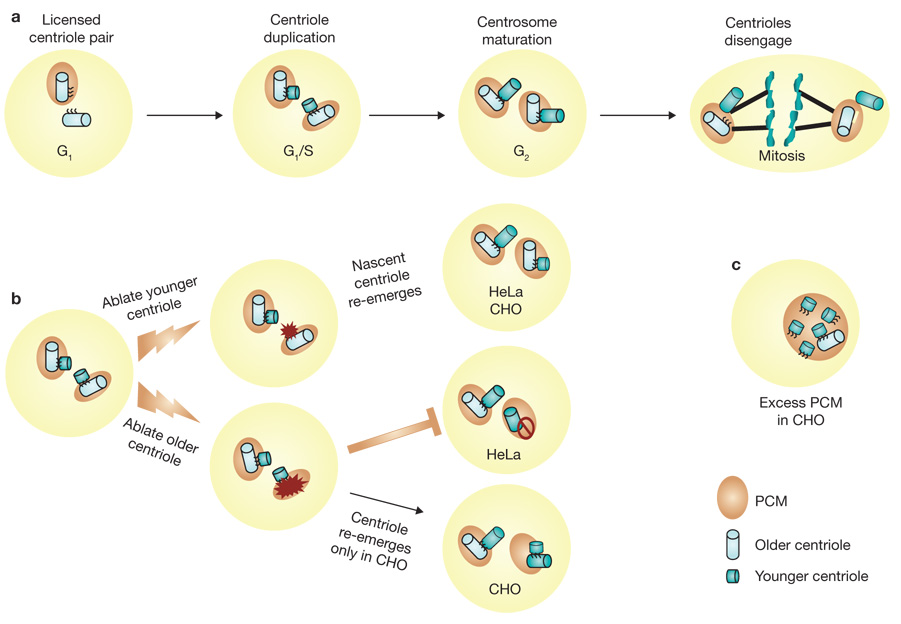
Alternative routes for centriole duplication. (a) Centrosomes of metazoan cells, including those of humans, consist of a pair of centrioles and a surrounding protein lattice called pericentriolar material (PCM), which serves to secure γ-tubulin-containing microtubule nucleation sites. As cells enter S phase, new centrioles begin to form near each of the two pre-existing centrioles and establish an orthogonal arrangement for each centriole pair with the nascent centriole extending from the proximal wall of the older centriole. During late S and G2 phases of the cell cycle, the newly formed centrioles elongate to near mature length. As cells prepare for mitosis, the centrosome matures by acquiring additional PCM and γ-tubulin, and then the two pairs of centrioles and their associated PCM separate to function as two mitotic spindle poles of the dividing cell. As cells pass through anaphase, the individual centrioles of each pair separate a short distance from one another in a process called centriole disengagement. Finally, with the completion of cell division, each G1 daughter cell inherits one spindle pole (centrosome) that contains a disoriented pair of centrioles. (b) Laser ablation studies show that when the younger centriole of a pair is destroyed, a new centriole emerges within several hours in both HeLa and CHO cells. However, when the older centriole of a pair is destroyed, a new centriole re-emerges only in CHO cells. (c) CHO cells expressing excess pericentrin form multiple nascent centrioles.
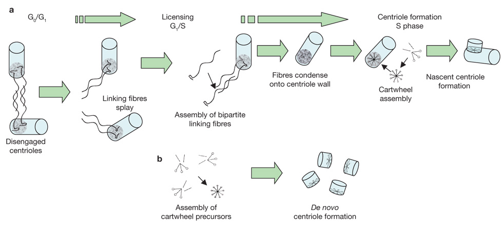
A unifying model for control of centriole number. (a) Semi-conservative centriole duplication: cells are produced in G0/G1 with a pair of disengaged centrioles. Licensing centriole duplication involves the dissociation (splaying) of linking fibre arrays, half of which remain attached to each pre-existing centriole. This is followed by formation of a complete ‘unit of duplication’ through the assembly of new bipartite fibre arrays. The fibres then contract or condense on the proximal wall of the centriole to which they are attached and form the preferred site for assembly of the cartwheel structure. The cartwheel then becomes a template for formation of a nascent centriole. In this manner, strict control over centriole number is maintained by the older centriole. (b) De novo centriole formation: in some cells, and under exceptional experimental conditions, new centrioles can form when the concentration of precursor molecules becomes high enough to cause cartwheel formation despite the lack of a favoured assembly site. De novo centriole formation does not exercise strict control over centriole number.
Comment on
-
Control of daughter centriole formation by the pericentriolar material.Nat Cell Biol. 2008 Mar;10(3):322-8. doi: 10.1038/ncb1694. Epub 2008 Feb 24. Nat Cell Biol. 2008. PMID: 18297061 Free PMC article.
References
Publication types
MeSH terms
Substances
Grants and funding
LinkOut - more resources
Full Text Sources
