. 2008 Nov;57(11):1665-73.
doi: 10.1007/s00262-008-0487-4. Epub 2008 Mar 1.

Inefficient presentation of tumor-derived antigen by tumor-infiltrating dendritic cells

Affiliations

Inefficient presentation of tumor-derived antigen by tumor-infiltrating dendritic cells

Patrizia Stoitzner et al. Cancer Immunol Immunother. 2008 Nov.

Abstract

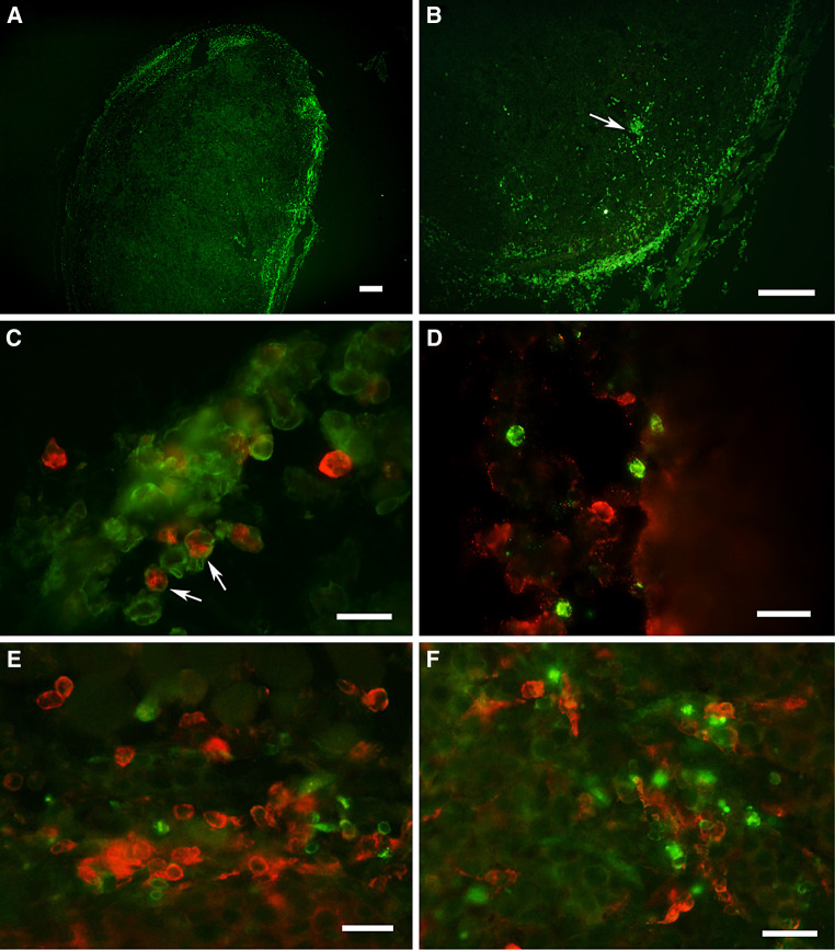
Background: Transplantable B16 melanoma is widely used as a tumor model to investigate tumor immunity. We wished to characterize the leukocyte populations infiltrating B16 melanoma tumors, and the functional properties of tumor-infiltrating dendritic cells (TIDC).

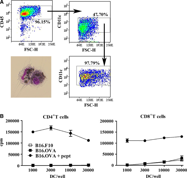
Materials and methods: We used the B16 melanoma cell line expressing ovalbumin protein (OVA) to investigate the phenotype and T cell stimulatory capacity of TIDC.

Results: The majority of leukocytes in B16 melanoma were macrophages, which colocalized with TIDCs, B and T cells to the peripheral area of the tumor. Both myeloid and plasmacytoid DC populations were present within tumors. Most of these DCs appeared immature, but about a third expressed a mature phenotype. TIDCs did not present tumor-derived antigen, as they were unable to induce the proliferation of tumor-specific CD4+ and CD8+ T cells in vitro unless in the presence of specific peptides. Some presentation of tumor-derived antigen could be demonstrated in the tumor-draining lymph node using in vivo proliferation assays. However, while proliferation of CD8+ T cells was reproducibly demonstrated, no proliferation of CD4+ T cells was observed.

Conclusion: In summary, our data suggest that DCs in tumors have limited antigen-presenting function. Inefficient antigen presentation extends to the tumor-draining lymph node, and may affect the generation of antitumor immune responses.


Figures

Fig. 1
Fig. 1
Characterization of leukocyte infiltrate in tumors and draining lymph nodes in melanoma-bearing mice. Mice were injected s.c. into the flank with 105 B16.OVA tumor cells. After 10–14 days, tumors had reached a size of 0.5–1.5 cm2 and were excised, digested with collagenase and analyzed by flow cytometry. a CD45+ leukocytes in tumor cell suspensions were gated, and percentages of different cell populations are shown as mean ± SD (summary of five experiments). b Percent expression of lineage and activation markers on CD11c+ TIDCs is shown as mean ± SD (summary of three different experiments). c Tumor-draining and contralateral lymph nodes were excised from mice carrying B16.OVA tumors and compared to control lymph nodes. Cell suspensions were obtained by digestion with collagenase and analyzed by flow cytometry. Total numbers of different cell populations are summarized from four different experiments and are shown as mean ± SD
Fig. 2
Fig. 2
Leukocytes are located in the periphery of tumors. Mice were injected s.c. into the flank with 105 B16.OVA tumor cells. Frozen sections from tumors (size 0.5–0.8 cm2) were prepared. Tumor sections were stained with mAb specific for macrophages, DCs, B and T cells, and viewed on a conventional immunofluorescence microscope. a, b Tumor sections stained with anti-CD45 mAb. CD45+ leukocytes are located in the periphery of the tumor. The arrow indicates leukocytes close to blood vessel. Bars in a and b represent 200 μm. c Double staining for CD11c (green fluorescence) and MHC II (red fluorescence). CD11c+ DCs express MHC-class II mostly inside the cells indicating an immature phenotype. MHC-class II-single positive cells could be either macrophages or B cells. d Double staining for CD11c+ DCs (green fluorescence) and B220+ B cells (red fluorescence). e Colocalization of CD4+ T cells (green fluorescence) and CD11c+ DC (red fluorescence) to the same area. f Colocalization of CD8+ T cells (green fluorescence) and CD11c+ DC (red fluorescence). Bars in cf correspond to 20 μm
Fig. 3
Fig. 3
TIDCs cannot present tumor-derived antigen in vitro. We injected mice with B16.OVA or control B16.F10, excised the tumors after 14 days (size of tumors 0.5–1.5 cm2) and digested them with collagenase. Cell suspensions were pre-enriched for CD45+ cells by magnetic bead sorting and electronically sorted for CD11c+ DC. a The sorting strategy and purity of TIDCs are shown. TIDCs contain melanin that can be seen as black particles inside the sorted DCs. b Graded doses of tumor-derived DC were cocultured with 2 × 105 OVA-specific CD4+ OT-II and CD8+ OT-I T cells for 3 days, and radioactive thymidine was added during the last 16 h. Results are expressed as counts per minute (cpm). Cultures were set up in triplicate; mean ± SD are shown. Results are from one of four experiments that gave similar results
Fig. 4
Fig. 4
Tumor-derived antigen is presented to CD8+ T cells in tumor-draining lymph nodes in vivo. CD45.1+ congenic mice were injected subcutaneously into one flank with 105 B16.OVA or control B16.F1 tumor cells. Ten days later, CFSE-labelled OVA-specific CD4+ OT-II and CD8+ OT-I T cells were adoptively transferred into tumor-bearing mice. The tumor-draining and contralateral nondraining lymph nodes were examined for the presence of proliferating T cells 3 days later. a shows complete data for all mice in one experiment are shown. The percent proliferating T cells was determined as shown in b. b Histograms for CFSE-proliferation of CD45.1+ cells from one representative mouse from each group are shown. Each symbol represents one mouse; means are indicated by horizontal bars. Data are from one of three experiments that gave similar results

References

    1. Barnden MJ, Allison J, Heath WR, Carbone FR. Defective TCR expression in transgenic mice constructed using cDNA-based alpha- and beta-chain genes under the control of heterologous regulatory elements. Immunol Cell Biol. 1998;76:34–40. doi: 10.1046/j.1440-1711.1998.00709.x. - DOI - PubMed
    1. Bell D, Chomarat P, Broyles D, Netto G, Harb GM, Lebecque S, Valladeau J, Davoust J, Palucka KA, Banchereau J. In breast carcinoma tissue, immature dendritic cells reside within the tumor, whereas mature dendritic cells are located in peritumoral areas. J Exp Med. 1999;190:1417–1426. doi: 10.1084/jem.190.10.1417. - DOI - PMC - PubMed
    1. Bennaceur K, Popa I, Portoukalian J, Berthier-Vergnes O, Peguet-Navarro J. Melanoma-derived gangliosides impair migratory and antigen-presenting function of human epidermal Langerhans cells and induce their apoptosis. Int Immunol. 2006;18:879–886. doi: 10.1093/intimm/dxl024. - DOI - PubMed
    1. Berthier-Vergnes O, Gaucherand M, Peguet-Navarro J, Plouet J, Pageaux JF, Schmitt D, Staquet MJ. Human melanoma cells inhibit the earliest differentiation steps of human Langerhans cell precursors but failed to affect the functional maturation of epidermal Langerhans cells. Br J Cancer. 2001;85:1944–1951. doi: 10.1054/bjoc.2001.2183. - DOI - PMC - PubMed
    1. Chiodoni C, Paglia P, Stoppacciaro A, Rodolfo M, Parenza M, Colombo MP. Dendritic cells infiltrating tumors cotransduced with granulocyte/macrophage colony-stimulating factor (GM-CSF) and CD40 ligand genes take up and present endogenous tumor-associated antigens, and prime naive mice for a cytotoxic T lymphocyte response. J Exp Med. 1999;190:125–133. doi: 10.1084/jem.190.1.125. - DOI - PMC - PubMed

Publication types

MeSH terms