. 2008 Apr;68(1):37-50.
doi: 10.1111/j.1365-2958.2008.06131.x. Epub 2008 Feb 26.

Characterization of Trypanosoma brucei dihydroorotate dehydrogenase as a possible drug target; structural, kinetic and RNAi studies

Affiliations

Characterization of Trypanosoma brucei dihydroorotate dehydrogenase as a possible drug target; structural, kinetic and RNAi studies

Tracy L Arakaki et al. Mol Microbiol. 2008 Apr.

Abstract

Nucleotide biosynthesis pathways have been reported to be essential in some protozoan pathogens. Hence, we evaluated the essentiality of one enzyme in the pyrimidine biosynthetic pathway, dihydroorotate dehydrogenase (DHODH) from the eukaryotic parasite Trypanosoma brucei through gene knockdown studies. RNAi knockdown of DHODH expression in bloodstream form T. brucei did not inhibit growth in normal medium, but profoundly retarded growth in pyrimidine-depleted media or in the presence of the known pyrimidine uptake antagonist 5-fluorouracil (5-FU). These results have significant implications for the development of therapeutics to combat T. brucei infection. Specifically, a combination therapy including a T. brucei-specific DHODH inhibitor plus 5-FU may prove to be an effective therapeutic strategy. We also show that this trypanosomal enzyme is inhibited by known inhibitors of bacterial Class 1A DHODH, in distinction to the sensitivity of DHODH from human and other higher eukaryotes. This selectivity is supported by the crystal structure of the T. brucei enzyme, which is reported here at a resolution of 1.95 A. Additional research, guided by the crystal structure described herein, is needed to identify potent inhibitors of T. brucei DHODH.


Figures

Figure 1
Figure 1
Oxidation of dihydroorotate to orotate.
Figure 2
Figure 2
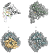
Comparison of T. brucei and human DHODH. TbDHODH is homodimeric, typical of Class 1A homologues. The human enzyme is monomeric, characteristic of Class 2 homologues. (a) TbDHODH monomer showing Class 1A motifs: Motif 1 (residues 195–203, blue), Motif 2 (residues 64–76, green), Motif 3 (residues 214–223, orange), Pyr-binding motif (residues 126–143, red). The orotate and FMN cofactors are shown in brown and yellow, respectively. (b) Surface representation of TbDHODH, showing that the orotate and FMN sites are relatively solvent accessible even with loop 132–138 in a ‘closed’ conformation. (c) Surface representation of the entire human DHODH monomer including well-ordered residues (30–69) from the N-terminal domain (brown). In Class 2 homologues the N-terminal domain restricts solvent access to the orotate and FMN binding sites from this direction. The depression visible at the center of the surface is the binding site for the Class 2 DHODH redox partner ubiquinone, and for Class 2 specific inhibitors such as brequinar. (d) Surface representation the human DHODH with N-terminal residues removed for the purpose of comparison to the trypanosomal enzyme.
Figure 3
Figure 3
A structure-based sequence alignment of the Class 1A T. brucei (current structure), the Class 1B L. lactis (PDB accession code 1ep1), and the Class 2 human DHODH (PDB accession code 1d3g). The active site Cys131 is colored yellow. Four Asn residues important for catalysis and conserved in all classes of DHODH are colored cyan. Three Class 1A motifs that may further aid in the classification of DHODHs are labeled and further discussed in Fig. S2. Residue numbering and secondary structure elements correspond to the T. brucei DHODH. The alignment was generated using the CEMC Multiple Protein Structure Alignment Server (http://bioinformatics.albany.edu/~cemc/) (Guda et al., 2004) and colored using TEXshade (Beitz, 2000). The human sequence shown begins at residue 69, omitting the N-terminal domain that is not present in the Class 1 homologues.
Figure 4
Figure 4
Knockdown of DHODH mRNA expression (Panel A) and DHODH protein expression (Panel B) after induction of RNAi with tetracycline. (A - top) Northern blot of T. brucei cells collected 72 h post-tetracycline induction of RNAi (TCN+) demonstrates 95% knockdown of DHODH mRNA compared to cells containing the same construct with RNAi uninduced (TCN-). (A - bottom) β-tubulin mRNA expression levels show near-equivalent RNA loading. (B) A Western blot was performed on cell lysates collected on day 0 (pre-induction) through 7 days post-induction. The blot was probed with an anti-T. brucei DHODH antibody. The upper band represents binding to an unidentified protein that is not affected by the RNAi expression. The predicted DHODH protein is labelled and is nearly absent one day after induction of RNAi.
Figure 5
Figure 5
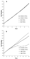
Gene knockdown of T. brucei DHODH using RNA interference. Cumulative cell densities are shown on a log-scale as the product of the cell number and the total dilution. (A). Cell growth was monitored of T. brucei undergoing RNAi of DHODH (TCN+) and compared to cells without RNAi induction (TCN−). The cells were grown in standard media for bloodstream-form T. brucei. The growth rate was also compared to the "single marker" (SM) strain of T. brucei that was not transfected with the RNAi expression vector (semi-logarithmic plot). (B). The same experiment as in Panel A was repeated using pyrimidine-depleted medium.
Figure 6
Figure 6
Effects of different concentrations of 5-FU on T. brucei cells. RNAi was induced with tetracycline for 48 h, then cells were incubated with serial dilutions of 5-FU for another 48 h before being quantified with Alamar Blue. Labeling is the same as in Figure 5.
Figure 7
Figure 7
Incorporation of (3H)5-FU into T. brucei cells. RNAi was induced with tetracycline for 72 h, then cells were exposed to (3H)5-FU for an additional 24 h before harvesting and quantifying tritium uptake into DNA. Labeling is the same as in Figure 5.
Figure 8
Figure 8
Terminal electron acceptor preference of TbDHODH. Activity was examined in the presence of 250 µM DHO and 100 µM electron acceptor or ~210 µM molecular oxygen. The kcatapp is the apparent kcat obtained at a fixed concentration of both DHO (0.25 mM) and oxidant.
Figure 9
Figure 9
Saturation kinetics of T. brucei DHODH toward DHO and fumarate. DHO-dependent activity was assayed in the presence of 500 µM fumarate and fumarate-dependent activity was assayed in the presence of 250 µM DHO at pH 8. Vmax is 8.5 s−1, Km for DHO is 14 µM, Km for fumarate is 80 µM. The kcatapp is the apparent kcat obtained at a fixed concentration of one substrate while the other is varied. Inset: pH-dependent T. brucei DHODH activity. Activity was determined in the presence of 125 µM DHO and 500 µM fumarate. Activity peaks at pH 7.8 and the curve indicates two ionizable groups with pKa values of 6.3 and 9.2.
Figure 10
Figure 10
Ping-pong kinetics of T. brucei DHODH. Double-reciprocal plot of DHO-dependent activity in the presence of varying concentrations of fumarate and 5 nM T. brucei DHODH produce parallel lines indicative of a ping-pong enzyme mechanism.
Figure 11
Figure 11
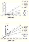
Inhibition of TbDHODH by 3,4- or 3,5-dihydroxybenzoate. Activity was determined at varying concentrations of DHO and inhibitor and 5 nM T. brucei DHODH. Double reciprocal plots show characteristic competitive inhibition by the indicated concentrations 3,4 dihydroxybenzoate (a) and non-competitive inhibition by 3,5 dihydroxybenzoate (b).

References

    1. Alexandrov A, Vignali M, LaCount DJ, Quartley E, de Vries C, Rosa DD, Babulski J, Mitchell SF, Schoenfeld LW, Fields S, Hol WG, Dumont ME, Phizicky EM, Grayhack EJ. A facile method for high-throughput co-expression of protein pairs. Mol Cell Proteomics. 2004;3:934–938. - PubMed
    1. Andersen PS, Jansen PJ, Hammer K. Two different dihydroorotate dehydrogenases in Lactococcus lactis. J Bacteriol. 1994;176:3975–3982. - PMC - PubMed
    1. Annoura T, Nara T, Makiuchi T, Hashimoto T, Aoki T. The origin of dihydroorotate dehydrogenase genes of kinetoplastids, with special reference to their biological significance and adaptation to anaerobic, parasitic conditions. J Mol Evol. 2005;60:113–127. - PubMed
    1. Baldwin J, Michnoff CH, Malmquist NA, White J, Roth MG, Rathod PK, Phillips MA. High-throughput screening for potent and selective inhibitors of Plasmodium falciparum dihy-droorotate dehydrogenase. J Biol Chem. 2005;280:21847–21853. - PubMed
    1. Beitz E. TEXshade: shading and labeling of multiple sequence alignments using LATEX2 epsilon. Bioinformatics. 2000;16:135–139. - PubMed

Publication types

MeSH terms

LinkOut - more resources