. 2008 Feb 29:7:10.
doi: 10.1186/1475-925X-7-10.

Mathematical modeling of laser lipolysis

Affiliations

Mathematical modeling of laser lipolysis

Serge R Mordon et al. Biomed Eng Online. .

Abstract

Background and objectives: Liposuction continues to be one of the most popular procedures performed in cosmetic surgery. As the public's demand for body contouring continues, laser lipolysis has been proposed to improve results, minimize risk, optimize patient comfort, and reduce the recovery period. Mathematical modeling of laser lipolysis could provide a better understanding of the laser lipolysis process and could determine the optimal dosage as a function of fat volume to be removed.

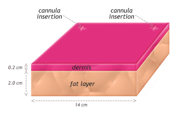
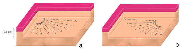
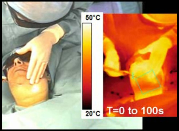
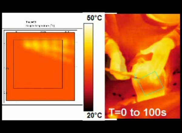
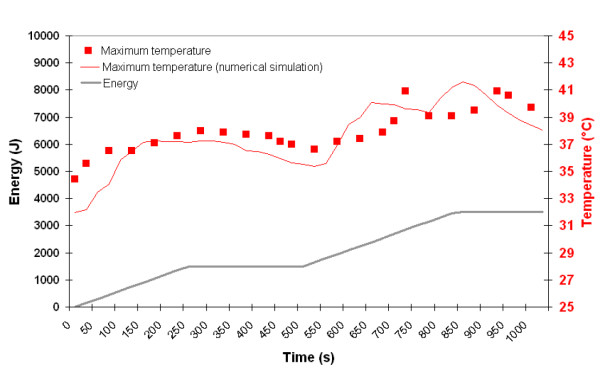
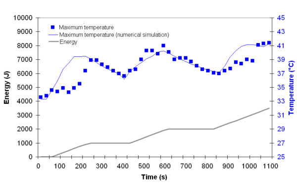
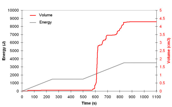
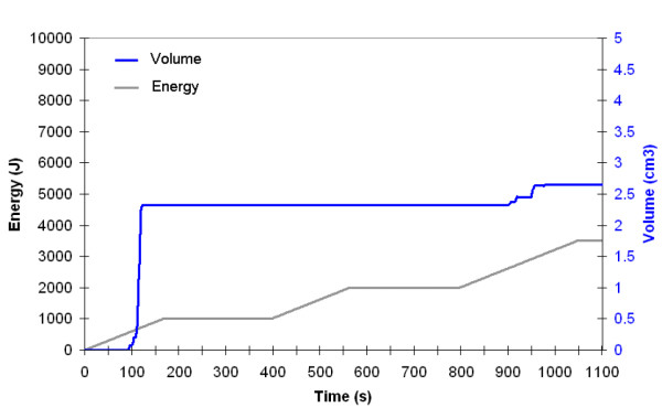
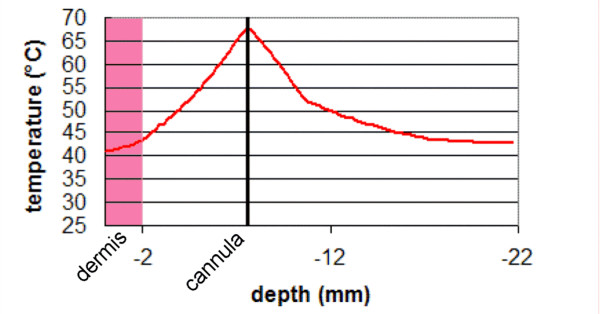
Study design/materials and methods: An Optical-Thermal-Damage Model was formulated using finite-element modeling software (Femlab 3.1, Comsol Inc). The general model simulated light distribution using the diffusion approximation of the transport theory, temperature rise using the bioheat equation and laser-induced injury using the Arrhenius damage model. Biological tissue was represented by two homogenous regions (dermis and fat layer) with a nonlinear air-tissue boundary condition including free convection. Video recordings were used to gain a better understanding of the back and forth movement of the cannula during laser lipolysis in order to consider them in our mathematical model. Infrared video recordings were also performed in order to compare the actual surface temperatures to our calculations. The reduction in fat volume was determined as a function of the total applied energy and subsequently compared to clinical data reported in the literature.

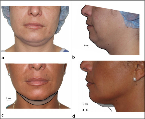
Results: In patients, when using cooled tumescent anesthesia, 1064 nm Nd:YAG laser or 980 nm diode laser: (6 W, back and forth motion: 100 mm/s) give similar skin surface temperature (max: 41 degrees C). These measurements are in accordance with those obtained by mathematical modeling performed with a 1 mm cannula inserted inside the hypodermis layer at 0.8 cm below the surface. Similarly, the fat volume reduction observed in patients at 6-month follow up can be determined by mathematical modeling. This fat reduction depends on the applied energy, typically 5 cm3 for 3000 J. At last, skin retraction was observed in patients at 6-month follow up. This observation can be easily explained by mathematical modeling showing that the temperature increase inside the lower dermis is sufficient (48-50 degrees C) to induce skin tightening

Discussion and conclusion: Laser lipolysis can be described by a theoretical model. Fat volume reduction observed in patients is in accordance with model calculations. Due to heat diffusion, temperature elevation is also produced inside the lower reticular dermis. This interesting observation can explain remodeling of the collagenous tissue, with clinically evident skin tightening. In conclusion, while the heat generated by interstitial laser irradiation provides stimulate lipolysis of the fat cells, the collagen and elastin are also stimulated resulting in a tightening in the skin. This mathematical model should serve as a useful tool to simulate and better understand the mechanism of action of the laser lipolysis.


Figures

Figure 1
Figure 1
Model geometry: 3D model consisting of a volume of tissue with two different layers: a dermal layer (thickness: 2 mm) and a hypodermis layer (thickness: 20 mm). The dimensions of this volume were 14 cm × 14 cm × 2.2 cm. This volume is s surrounded by infinite and homogenous tissue
Figure 2
Figure 2
a: Model geometry: The cannula is inserted inside the hypodermis layer at approximately 0.8 cm below the surface. This cannula is moved back and forth onto 100 mm in the fat layer with a velocity of 100 mm/in a plane parallel to the surface. This back and forth motion is repeated 15 times for each position of the cannula. This step is repeated 9 times (10 degrees angle each time). 2b: in order to provide a homogeneous treatment of the fat layer, the cannula is inserted at a distance of 120 mm from the previous insertion point and the procedure is repeated.
Figure 3
Figure 3
video recording of a laser lipolysis procedure inside the submentum. Conventional and infrared video recordings are performed simultaneously.
Figure 4
Figure 4
infrared video recording of a laser lipolysis procedure inside the submentum (right) and mathematical modeling performed of a specific area (green square of submentum of the patient).
Figure 5
Figure 5
Maximum temperature recorded on skin surface (red square) using the infrared camera and determined at the same position by numerical simulation (red line) as a function time and energy. The energy delivery as function of time is traced (gray line). Right side: 980 nm diode laser, power: 6W, CW, back and forth motion: 100 mm/s).
Figure 6
Figure 6
Maximum temperature recorded on skin surface (blue square) using the infrared camera and determined at the same position by numerical simulation (blue line) as a function time and energy The energy delivery as function of time is traced (gray line). Left side: 1064 nm Nd:YAG laser, power: 6W, CW, back and forth motion: 100 mm/s).
Figure 7
Figure 7
Volume reduction (red line) determined by numerical simulation as a function time and energy (980 nm-diode laser). The energy delivery as function of time is traced (gray line). Right side: 980 nm diode laser, power: 6W, CW, back and forth motion: 100 mm/s.
Figure 8
Figure 8
Volume reduction (blue line) determined by numerical simulation as a function time and energy. The energy delivery as function of time is traced (gray line). Left side: 1064 nm Nd:YAG laser, power: 6W, CW, back and forth motion: 100 mm/s).
Figure 9
Figure 9
shows pictures of the patient before and at 6-month follow-up. - a and b: before laser lipolysis, - c and d: 6 months after laser lipolysis. Right side: 980 nm diode laser, power: 6W, CW, back and forth motion: 100 mm/s, total energy: 3500 J). Left side: 1064 nm Nd:YAG laser, power: 6W, CW, back and forth motion: 100 mm/s, total energy: 3100 J)
Figure 10
Figure 10
maximum temperature elevation recorded inside the skin during laser lipolysis. Parameters: 980 nm diode laser, power: 6W, CW, back and forth motion: 100 mm/s, total energy: 3500 J.

References

    1. Apfelberg DB, Rosenthal S, Hunstad JP, Achauer B, Fodor PB. Progress report on multicenter study of laser-assisted liposuction. Aesthetic Plast Surg. 1994;18:259–264. doi: 10.1007/BF00449791. - DOI - PubMed
    1. Prado A, Andrades P, Danilla S, Leniz P, Castillo P, Gaete F. A prospective, randomized, double-blind, controlled clinical trial comparing laser-assisted lipoplasty with suction-assisted lipoplasty. Plast Reconstr Surg. 2006;118:1032–1045. doi: 10.1097/01.prs.0000232428.37926.48. - DOI - PubMed
    1. Goldman A. Submental Nd:Yag laser-assisted liposuction. Lasers Surg Med. 2006;38:181–184. doi: 10.1002/lsm.20270. - DOI - PubMed
    1. Badin AZ, Gondek LB, Garcia MJ, Valle LC, Flizikowski FB, de Noronha L. Analysis of laser lipolysis effects on human tissue samples obtained from liposuction. Aesthetic Plast Surg. 2005;29:281–286. doi: 10.1007/s00266-004-0102-9. - DOI - PubMed
    1. Mordon S, Eymard-Maurin AF, Wassmer B, Ringot J. Histologic evaluation of laser lipolysis: Pulsed 1,064nm Nd: YAG laser versus CW 980nm diode laser. Aesthetic Surgery Journal. 2007;27:263–268. doi: 10.1016/j.asj.2007.03.005. - DOI - PubMed