. 2008;10(1):R21.
doi: 10.1186/bcr1872. Epub 2008 Feb 29.

Forkhead box transcription factor FOXO3a suppresses estrogen-dependent breast cancer cell proliferation and tumorigenesis

Affiliations

Forkhead box transcription factor FOXO3a suppresses estrogen-dependent breast cancer cell proliferation and tumorigenesis

Yiyu Zou et al. Breast Cancer Res. 2008.

Abstract

Introduction: Estrogen receptors (ERs) play key roles in breast cancer development and influence treatment outcome in breast cancer patients. Identification of molecules that regulate ER function may facilitate development of breast cancer treatment strategies. The forkhead box class O (FOXO) transcription factor FOXO3a has been suggested to function as a tumor suppressor in breast cancer. Using protein-protein interaction screening, we found that FOXO3a interacted with ER-alpha and ER-beta proteins in the human breast carcinoma cell line MCF-7, suggesting that there exists a crosstalk between the FOXO3a and ER signaling pathways in estrogen-dependent breast cancer cells.

Methods: The interaction between FOXO3a and ER was investigated by using co-immunoprecipitation and immunoblotting assays. Inhibition of ER-alpha and ER-beta transactivation activity by FOXO was determined by luciferase reporter assays. Cell proliferation in culture was evaluated by counting cell numbers. Tumorigenesis was assessed in athymic mice that were injected with MCF-7 cell lines over-expressing FOXO3a. Protein expression levels of cyclin-dependent kinase inhibitors, cyclins, ERs, FOXM1, and the proteins encoded by ER-regulated genes in MCF-7 cell lines and breast tumors were examined by immunoblotting analysis and immunohistochemical staining.

Results: We found that FOXO3a interacted with ER-alpha and ER-beta proteins and inhibited 17beta-estradiol (E2)-dependent, ER-regulated transcriptional activities. Consistent with these observations, expression of FOXO3a in the ER-positive MCF-7 cells decreased the expression of several ER-regulated genes, some of which play important roles in cell proliferation. Moreover, we found that FOXO3a upregulated the expression of the cyclin-dependent kinase inhibitors p21Cip1, p27Kip1, and p57Kip2. These findings suggest that FOXO3a induces cell growth arrest to effect tumor suppression. FOXO3a repressed the growth and survival of MCF-7 cells in cell culture. In an orthotopic breast cancer xenograft model in athymic mice, over-expression of FOXO3a in MCF-7 cells suppressed their E2-induced tumorigenesis, whereas knockdown of FOXO3a in MCF-7 resulted in the E2-independent growth.

Conclusion: Functional interaction between FOXO3a and ER plays a critical role in suppressing estrogen-dependent breast cancer cell growth and tumorigenesis in vivo. This suggests that agents that activate FOXO3a may be novel therapeutic agents that can inhibit and prevent tumor proliferation and development in breast cancer.


Figures

Figure 1
Figure 1
FOXO3a interacts with ER-α and ER-β. (a) Total lysates of 293T cells co-transfected with hemagglutinin (HA)-tagged forkhead box class O (FOXO)3a plus Flag-estrogen receptor (ER)-α (lanes 2, 3) or an empty vector (lane 1) or an ErbB2 expression vector (lane 4), in the presence or absence of 17β-estradiol (E2), were analyzed by immunoprecipitation (IP) with an anti-HA antibody followed by immunoblotting (IB) with an anti-Flag tag antibody. (b) The same lysates of 293T cells cotransfected with HA-FOXO3a and Flag-ER-α were subjected to reciprocal IP with an anti-Flag (lane 3) or control IgG (IgG; lane 2) or anti-HA (positive control, lane 4) followed by IB with an anti-HA. (c) FOXO3a associates with ER-β. Total lysates of 293T cells co-transfected with the Flag-FOXO3a and HA-ER-β expression vectors in the presence of E2 were analyzed by the same IP/IB analysis (IP: control IgG or anti-Flag [positive control] or anti-HA [lane 3]; IB: anti-Flag). (d) The same lysates of 293T cells co-transfected with the Flag-FOXO3a and HA-ER-β expression vectors were subjected to reciprocal IP with a control IgG or anti-Flag (lane 2) or anti-HA (positive control) followed by IB with an anti-HA.
Figure 2
Figure 2
FOXO3a associates with endogenous ER-α and ER-β. (a, b) Forkhead box class O (FOXO)3a interacts with endogenous ER-α. Panel a: total lysates of MCF-7 cells with 17β-estradiol (E2) were subjected to immunoprecipitation (IP) with an anti-FOXO3a (positive control) or negative control IgGs (lanes 2 and 3) or anti-estrogen receptor (ER)-α (lane 4), followed by immunoblotting (IB) with an anti-FOXO3a antibody. Panel b: the same lysates of MCF-7 cells with E2 were subjected to reciprocal IP with an anti-FOXO3a antibody (lane 1) or a negative control IgG (lanes 2 and 3) or an anti-ER-α (positive control) followed by IB with an anti-ER-α. (c, d) FOXO3a interacts with endogenous ER-β. Panel c: total lysates of MCF-7 with E2 were subjected to IP/IB (IP: control IgG or anti-FOXO3a [positive control] or anti-ER-β (lane 4); IB: anti-FOXO3a). Panel d: total lysates of MCF-7 with E2 were subjected to a reciprocal IP/IB (IP: control IgG or anti-ER-β [positive control] or anti-FOXO3a [lane 4]; IB: anti-ER-β).
Figure 3
Figure 3
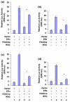
FOXO3a and FOXO1a inhibit the transactivation activities of ER-α and ER-β. (a, b) 293T cells were co-transfected with estrogen receptor (ER)-responsive element (ERE)-luc (firefly luciferase [luc] reporter containing EREs), pRL-TK (renilla luc as a transfection control for normalization), ER-α (panel a) or ER-β (panel b), and forkhead box class O (FOXO)3a plus IκB kinase (IKK)-β or an empty vector (control) as indicated. Total lysates of the transfected cells were prepared and subjected to luc assays. (c, d) Total lysates of 293T cells were co-transfected with ERE-luc, pRL-TK, ER-α (panel c) or ER-β (panel d), and FOXO1a plus IKK-β or an empty vector as indicated and subjected to luc assays. All cells were cultured in the presence of 17β-estradiol (E2). The relative reporter luc activity was normalized with pRL-TK. Data are expressed means and standard deviations from three repeated experiments, which were performed independently. *P < 0.05 between FOXO (FOXO3a or FOXO1a) minus IKK-β (lane 3) versus FOXO plus IKK-β (lane 4).
Figure 4
Figure 4
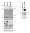
FOXO3a regulates expression of ER target genes and CDK inhibitors, and induces apoptosis in MCF-7. (a) Ectopic expression of Forkhead box class O (FOXO)3a reduces the expression of some estrogen receptor (ER)-regulated genes and enhances the expression of cyclin-dependent kinase (CDK) inhibitors in MCF7-FO cells in the presence of 17β-estradiol (E2). Immunoblotting (IB) analyses for HA-FOXO3a, endogenous FOXO3a, p27Kip1, p21Cip1, p57Kip2, cyclin D1, cyclin E, cathepsin D, progesterone receptor (PgR), ER-α, and ER-β protein expression in MCF7-FO33 and MCF7-FO41 cells (constitutively expressing FOXO3a) and in control (MCF-7 and MCF7-C5) cells were performed with specific antibodies, as indicated. Equal loading was confirmed by the same IB analysis with antibodies against β-actin or β-tubulin. (b) MDA-MB-453 (MDA-453, ER-negative) cells were transfected with either FOXO3a or an empty pCDNA3.1 vector (control), as indicated. Total lysates of the transfected cells were prepared and subjected to SDS-PAGE followed by IB analysis with the indicated antibodies.
Figure 5
Figure 5
Ectopic expression of FOXO3a in estrogen-dependent breast cancer cells suppresses cell proliferation in cell culture. (a) Growth of forkhead box class O (FOXO)3a over-expressing MCF7-FO (MCF7-FO10, MCF7-FO33, and MCF7-FO41) cells and control (MCF7-C4, MCF7-C5, and MCF7-C12) cells in the absence of 17β-estradiol (E2) was determined by counting trypan-blue stained cells with a hemocytometer. Growth curves are the means of the three MCF7-FO cell lines (MCF7-FO10, MCF7-FO33, and MCF7-FO41) and the three control cell lines (MCF7-C4, MCF7-C5, and MCF7-C12); error bars indicate standard deviation from three experiments. (b) Growth of the same sets of cells in the presence of E2 (1 nmol/l). Growth curves are the means of the three MCF7-FO cell lines (designated MCF7-FO average) and the three control cell lines (designated MCF7-C average); error bars indicate standard deviaton from three experiments. *P < 0.05 between control MCF7-C group versus MCF7-FO group.
Figure 6
Figure 6
Ectopic expression of FOXO3a in estrogen-dependent breast cancer cells suppresses breast tumor development in vivo. Forkhead box class O (FOXO)3a suppresses estrogen receptor (ER)-positive breast tumor development in a mouse model of breast cancer. The MCF7-FO pooled cell lines and the control MCF7-C pooled cell lines were injected (2 × 10⁶ cells/mouse) into the mammary fat pads of female athymic mice given supplementary 17β-estradiol (E2), as described in Materials and methods. Growth curves of tumor size are the means of the MCF7-FO pooled cell lines (designated MCF7-FO) and the control MCF7-C pooled cell lines (designated MCF7-C); error bars indicate standard deviation from three experiments. *P < 0.05 between control MCF7-C group versus MCF7-FO group.
Figure 7
Figure 7
FOXO3a decreases expression of ER-regulated genes and increases CDK inhibitors in MCF7-FO breast tumors. (a) At 35 days after tumor cell implantation, breast tumors derived from female athymic mice bearing MCF7-FO or MCF7-C (control) tumors were resected, fixed, sectioned, and placed on slides. Five independent tumors (each from a different mouse) were tested in each mouse group. Tumor specimens were subjected to immunohistochemical (IHC) staining with antibodies specific to forkhead box class O (FOXO)3a, hemagglutinin (HA)-tag, pS2, complement C3, cathepsin (Cath)-D, progesterone receptor (PgR), and cyclin D1. Slides were examined at 40× magnification with a microscope. Numbers of positively staining cells in four random fields were counted in each tumor section, and representative fields are shown. Scale bars indicate 50 μm. *P < 0.01 between control MCF7-C group versus MCF7-FO group. (b) At 35 days after tumor cell implantation, breast tumors derived from female athymic mice bearing MCF7-FO or MCF7-C (control) tumors were resected, fixed, sectioned, and placed on slides. Five independent tumors (each from a different mouse) were tested in each mouse group. Tumor specimens were subjected to IHC staining with antibodies to p21Cip1, p27Kip1, p57Kip2, estrogen receptor (ER)-α, and ER-β. Numbers of positively staining cells in four random fields were counted in each tumor section, and representative fields are shown. Scale bars indicate 50 μm. *P < 0.02 between control MCF7-C group versus MCF7-FO group. (c) Confirmation that FOXO3a enhances expression of cyclin-dependent kinase (CDK) inhibitors and reduces expression of cyclin D1 in tumors in immunoblotting (IB) analysis. Whole lysates of MCF7-FO or MCF7-C breast tumor specimens were subjected to IB analysis with antibodies to p21Cip1, p27Kip1, p57Kip2, cyclin D1, ER-α, ER-β, HA-tag and FOXO3a (positive controls), and β-actin (loading control).
Figure 8
Figure 8
ER-α and ER-β bind to the amino-terminal and carboxyl-terminal domains of FOXO3a, respectively. (a) Glutathione-S-transferase (GST) – pull down in vitro assays. Whole cell lysates from 293T cells were incubated with the GST-forkhead box class O (FOXO)3a (GST-FO [amino acids 1 to 300] and GST-FO [amino acids 301 to 673]) fusion proteins as indicated and GST alone (negative control), and analyzed by SDS-PAGE and immunoblotting with an antibody (Ab) against estrogen receptor (ER)-α or ER-β (upper panels) and an anti-GST Ab (lower panel) as protein controls. (b) FOXO3a downregulates FOXM1. Immunoblotting (IB) analyses for endogenous FOXO3a, forkhead box M1 (FOXM1), and β-actin (loading control) protein expression in MCF7-FO10 and MCF7-FO41 cells (constitutively expressing FOXO3a) and in control (MCF-7 wt and MCF7-C12) cells were performed with specific antibodies antibodies as indicated. (c) MCF7-d8_pa cells (pooled clones of MCF-7 FOXO3a-knockdown derivatives) were established with retroviruses expressing small hairpin RNA against human FOXO3a. The expression levels of FOXO3a and p27Kip1 in MCF-7 wild-type (wt) and MCF7-d8_pa cells were determined by IB with specific Abs against FOXO3a or p27Kip1 or β-actin (loading control). (d) Silencing endogenous FOXO3a in MCF-7 cells promoted tumorigenesis in vivo. The tumor growth rates of control group MCF-7 wt and knockdown group MCF7-d8-pa were determined after injection of cells (2 × 10⁶cells/mouse) as indicated into the mammary fat pads of female athymic mice not given supplemental 17β-estradiol (E2; indicated as – E2). Data are expressed as means and standard deviations from two experiments with five mice in each group. *P < 0.01 between MCF7-d8-pa group versus control MCF-7 wt group.

References

    1. Parkin DM, Bray F, Ferlay J, Pisani P. Global cancer statistics, 2002. CA Cancer J Clin. 2005;55:74–108. - PubMed
    1. Johnston SR, Dowsett M. Aromatase inhibitors for breast cancer: lessons from the laboratory. Nat Rev Cancer. 2003;3:821–831. doi: 10.1038/nrc1211. - DOI - PubMed
    1. Katzenellenbogen BS, Frasor J. Therapeutic targeting in the estrogen receptor hormonal pathway. Semin Oncol. 2004;31(Suppl 3):28–38. doi: 10.1053/j.seminoncol.2004.01.004. - DOI - PubMed
    1. Keen JC, Davidson NE. The biology of breast carcinoma. Cancer. 2003;97(Suppl):825–833. doi: 10.1002/cncr.11126. - DOI - PubMed
    1. Thiantanawat A, Long BJ, Brodie AM. Signaling pathways of apoptosis activated by aromatase inhibitors and antiestrogens. Cancer Res. 2003;63:8037–8050. - PubMed

Publication types