Lysosomal cholesterol accumulation inhibits subsequent hydrolysis of lipoprotein cholesteryl ester
- PMID: 18312718
- PMCID: PMC2837357
- DOI: 10.1017/S1431927608080069
Lysosomal cholesterol accumulation inhibits subsequent hydrolysis of lipoprotein cholesteryl ester
Abstract
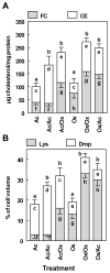
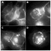
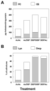
Human macrophages incubated for prolonged periods with mildly oxidized LDL (oxLDL) or cholesteryl ester-rich lipid dispersions (DISP) accumulate free and esterified cholesterol within large, swollen lysosomes similar to those in foam cells of atherosclerosis. The cholesteryl ester (CE) accumulation is, in part, the result of inhibition of lysosomal hydrolysis due to increased lysosomal pH mediated by excessive lysosomal free cholesterol (FC). To determine if the inhibition of hydrolysis was long lived and further define the extent of the lysosomal defect, we incubated THP-1 macrophages with oxLDL or DISP to produce lysosome sterol engorgement and then chased with acetylated LDL (acLDL). Unlike oxLDL or DISP, CE from acLDL normally is hydrolyzed rapidly. Three days of incubation with oxLDL or DISP produced an excess of CE in lipid-engorged lysosomes, indicative of inhibition. After prolonged oxLDL or DISP pretreatment, subsequent hydrolysis of acLDL CE was inhibited. Coincident with the inhibition, the lipid-engorged lysosomes failed to maintain an acidic pH during both the initial pretreatment and subsequent acLDL incubation. This indicates that the alterations in lysosomes were general, long lived, and affected subsequent lipoprotein metabolism. This same phenomenon, occurring within atherosclerotic foam cells, could significantly affect lesion progression.
Figures


Similar articles
-
Aggregated LDL and lipid dispersions induce lysosomal cholesteryl ester accumulation in macrophage foam cells.J Lipid Res. 2005 Oct;46(10):2052-60. doi: 10.1194/jlr.M500059-JLR200. Epub 2005 Jul 16. J Lipid Res. 2005. PMID: 16024919
-
Lysosomal cholesterol derived from mildly oxidized low density lipoprotein is resistant to efflux.J Lipid Res. 2001 Mar;42(3):317-27. J Lipid Res. 2001. PMID: 11254742
-
Cholesterol and oxysterol metabolism and subcellular distribution in macrophage foam cells. Accumulation of oxidized esters in lysosomes.J Lipid Res. 2000 Feb;41(2):226-37. J Lipid Res. 2000. PMID: 10681406
-
The role of microscopy in understanding atherosclerotic lysosomal lipid metabolism.Microsc Microanal. 2003 Feb;9(1):54-67. doi: 10.1017/S1431927603030010. Microsc Microanal. 2003. PMID: 12597787 Review.
-
Macrophage-mediated cholesterol handling in atherosclerosis.J Cell Mol Med. 2016 Jan;20(1):17-28. doi: 10.1111/jcmm.12689. Epub 2015 Oct 23. J Cell Mol Med. 2016. PMID: 26493158 Free PMC article. Review.
Cited by
-
Phenotypic, Metabolic, and Functional Characterization of Experimental Models of Foamy Macrophages: Toward Therapeutic Research in Atherosclerosis.Int J Mol Sci. 2024 Sep 21;25(18):10146. doi: 10.3390/ijms251810146. Int J Mol Sci. 2024. PMID: 39337629 Free PMC article.
-
Meningeal Foam Cells and Ependymal Cells in Axolotl Spinal Cord Regeneration.Front Immunol. 2019 Nov 1;10:2558. doi: 10.3389/fimmu.2019.02558. eCollection 2019. Front Immunol. 2019. PMID: 31736973 Free PMC article.
-
7-Ketocholesterol in disease and aging.Redox Biol. 2020 Jan;29:101380. doi: 10.1016/j.redox.2019.101380. Epub 2019 Nov 14. Redox Biol. 2020. PMID: 31926618 Free PMC article. Review.
-
Lysosomal acid lipase: at the crossroads of normal and atherogenic cholesterol metabolism.Front Cell Dev Biol. 2015 Feb 2;3:3. doi: 10.3389/fcell.2015.00003. eCollection 2015. Front Cell Dev Biol. 2015. PMID: 25699256 Free PMC article. Review.
-
Triglyceride alters lysosomal cholesterol ester metabolism in cholesteryl ester-laden macrophage foam cells.J Lipid Res. 2009 Oct;50(10):2014-26. doi: 10.1194/jlr.M800659-JLR200. Epub 2009 May 21. J Lipid Res. 2009. PMID: 19461120 Free PMC article.
References
-
- Auwerx J, Schoonjans K. New insights into apolipoprotein B and low density lipoprotein physiology; implications for atherosclerosis. Acta Clin Belg. 1991;46(6):355–8. - PubMed
-
- Barakat HA, St Clair RW. Characterization of plasma lipoproteins of grain- and cholesterol-fed white carneau and show racer pigeons. J Lipid Res. 1985;26:1252–1268. - PubMed
-
- Bligh EG, Dyer WJ. A rapid method for total lipid extraction and purification. Protein measurement with the Folin phenol reagent. Can J Biochem Physiol. 1959;37:911–917. - PubMed

