. 2008 Apr;82(4):809-21.
doi: 10.1016/j.ajhg.2008.01.010. Epub 2008 Feb 28.

Arrhythmogenic right ventricular cardiomyopathy type 5 is a fully penetrant, lethal arrhythmic disorder caused by a missense mutation in the TMEM43 gene

Affiliations

Arrhythmogenic right ventricular cardiomyopathy type 5 is a fully penetrant, lethal arrhythmic disorder caused by a missense mutation in the TMEM43 gene

Nancy D Merner et al. Am J Hum Genet. 2008 Apr.

Abstract

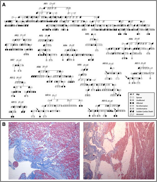
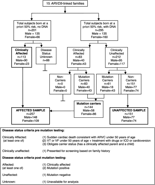
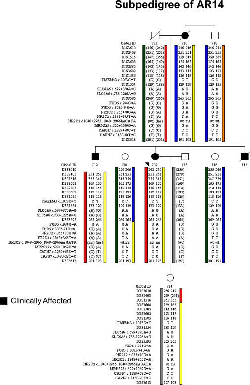
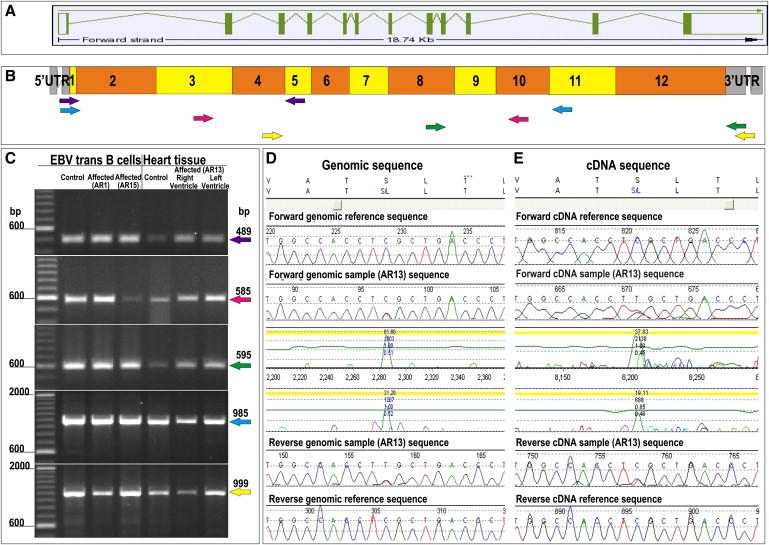
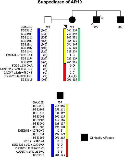
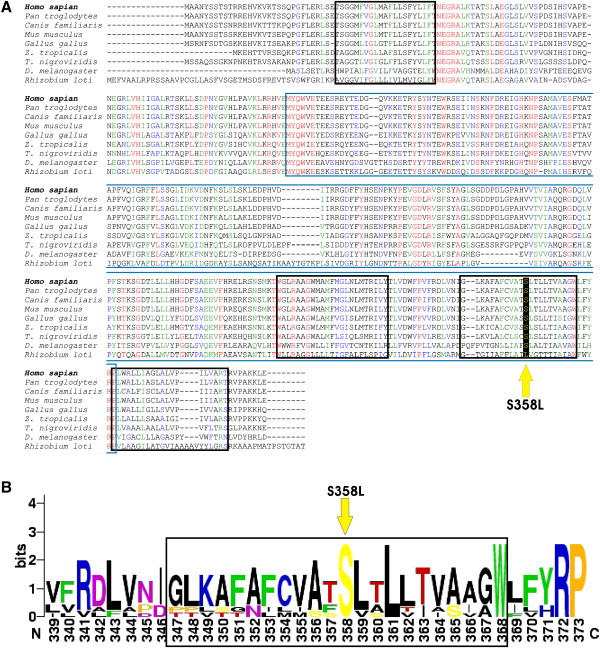
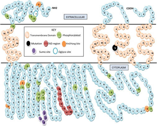
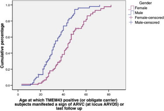
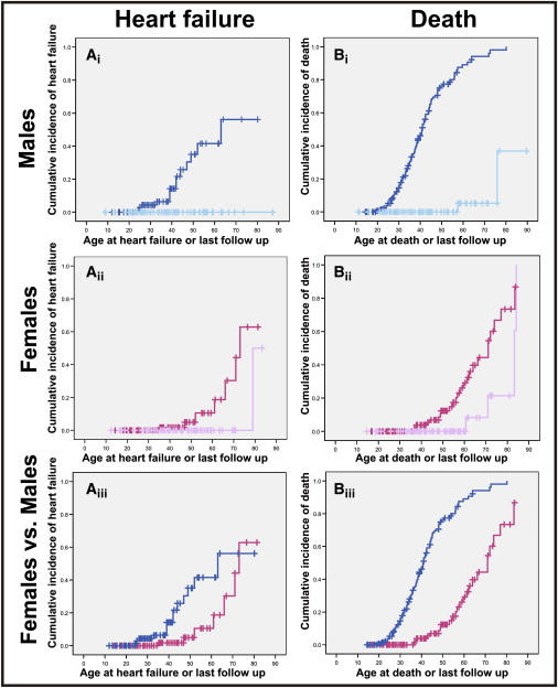
Autosomal-dominant arrhythmogenic right ventricular cardiomyopathy/dysplasia (ARVC/D) causes sudden cardiac death and is characterized by clinical and genetic heterogeneity. Fifteen unrelated ARVC families with a disease-associated haplotype on chromosome 3p (ARVD5) were ascertained from a genetically isolated population. Identification of key recombination events reduced the disease region to a 2.36 Mb interval containing 20 annotated genes. Bidirectional resequencing showed one rare variant in transmembrane protein 43 (TMEM43 1073C-->T, S358L), was carried on all recombinant ARVD5 ancestral haplotypes from affected subjects and not found in population controls. The mutation occurs in a highly conserved transmembrane domain of TMEM43 and is predicted to be deleterious. Clinical outcomes in 257 affected and 151 unaffected subjects were compared, and penetrance was determined. We concluded that ARVC at locus ARVD5 is a lethal, fully penetrant, sex-influenced morbid disorder. Median life expectancy was 41 years in affected males compared to 71 years in affected females (relative risk 6.8, 95% CI 1.3-10.9). Heart failure was a late manifestation in survivors. Although little is known about the function of the TMEM43 gene, it contains a response element for PPAR gamma (an adipogenic transcription factor), which may explain the fibrofatty replacement of the myocardium, a characteristic pathological finding in ARVC.


Figures

Figure 1
Figure 1
ARVC Families Linked to Chromosome 3p (A) Pedigrees of 15 autosomal-dominant ARVC families linked to ARVD5. Affected subjects are shown as blackened squares (male) and circles (female). Subjects deceased because of SCD are noted by a circle above the symbol. (B) Photomicrographs of paraffin-embedded postmortem right ventricular myocardium stained with masson trichrome showing fibrofatty replacement of myocytes from a male teenager after sudden cardiac death (left: 40× magnification) and a second-degree relative with sudden cardiac death in his eighth decade (right: 20× magnification). Pink represents normal myocardium, blue represents fiber, and white represents fat.
Figure 2
Figure 2
Workflow and Mutation Status of Subjects Born at a Priori 50% Risk of ARVC
Figure 3
Figure 3
Physical Map of the ARVD5 Critical Region (A) Summary recombinant ARVD5 haplotypes identified across the 15 ARVC families with microsatellite markers (alleles are either numbered [1–9] or given in base pairs). (B) The physical map of the ARVD5 critical region. Physical distances were captured from the March 2006 freeze of the UCSC Genome Browser. Arrows show the direction of transcription of each annotated gene.
Figure 4
Figure 4
Segregation Analysis in Subpedigree AR14 Of the 19 variants found exclusively in clinically affected subjects on the mutation-screening panel, only 11 were found to reside on the ARVD5 ancestral haplotype (yellow) through segregation analysis of clinically affected subjects (Global IDs 709, 710, and 712). Note that a clinically unaffected subject (Global ID 708) inherited a recombinant ARVD5 haplotype from her clinically affected mother that lacks TMEM43. Alleles in brackets have been inferred.
Figure 5
Figure 5
Gene Structure and Mutation Analysis of TMEM43 (A) Gene structure of TMEM43. Exons are represented by boxes. Translated exons are solid green, and untranslated exons are clear. Introns are represented by green lines. (B) Coverage of primers designed to amplify cDNA showing position of PCR primer pairs: exons 1–4 (purple), exons 4–9 (red), exons 9–12 (green), exons 1–10 (blue), and 5–12 (yellow). (C) PCR products amplified from cDNA of EBV-transfected B cells of affected subjects from the mutation-screening panel (affected AR1 and affected AR15) and unaffected (control) subjects. cDNA of heart tissue from both the left and right ventricle of an affected subject (AR13) and a heart biopsy from a control subject were size fractionated by electrophoresis. (D and E) Forward and reverse sequencing traces showing the TMEM43 1073C→T mutation of an affected subject's (AR13) genomic and cDNA. The amino acid translations (top) shows the S358L amino acid substitution.
Figure 6
Figure 6
Segregation Analysis in Subpedigree AR10 Clinically affected subjects (Global IDs 794 and 796) only have one of the five rare variants because of a historical recombination event on the ARVD5 haplotype (yellow). Alleles in brackets have been inferred.
Figure 7
Figure 7
Multiple Alignment of the TMEM43 Gene across Eight Eukaryotic and Prokaryotic Species (A) Clustal W align was used to align orthologs from Homo sapiens (NP_077310), Pan troglodytes (XP_516299), Canis familiaris (XP_541751), Mus musculus (NP_083042), Gallus gallus (XP_414378), Zenopus tropicalis (UP10004D5297), Tetraodon nigroviridis (Q4RXL8), Drosophila melanogaster (NP_64162), and Rhizobium loti (Q98HF3). The blue box outlines the DUF1625 domain, and the black boxes outline predicted transmembrane domains. Completely conserved residues are red, strongly similar residues are green, and weakly similar residues are blue. The S358L mutation is marked by a yellow arrow. (B) The web logo format was used for aligning eukaryotic species. The third transmembrane domain is outlined (black box). The S358L mutation is marked by a yellow arrow.
Figure 8
Figure 8
Predicted Topography of the TMEM43 Protein Indicated are transmembrane domains (beige), phosphorylation sites (green), a transactivation domain (red), YingOYang sites (orange), a SUMO attachment site (purple), and an O-glycosylation site (blue open). The extracellular and cytoplasmic regions may be reversed: There is evidence supporting either orientation.
Figure 9
Figure 9
ARVC Penetrance Caused by the TMEM43 1073C→T Mutation Cumulative proportion by age at which male (n = 60) and female (n = 77) mutation positive affected subjects manifested a first clinical sign of ARVC. Subjects were censored at last follow-up.
Figure 10
Figure 10
Time to Event Analysis of Heart Failure and Death in Affected Subjects (Ai–Aiii) Cumulative incidence of heart failure; panels describe the following: (Ai) affected males (n = 89, dark blue) compared to unaffected males (n = 71, light blue) (p ≤ 0.0001: log rank); (Aii) affected females (n = 87, dark pink) compared to unaffected females (n = 68, light pink) (p ≤ 0.001, log rank); and (Aiii) affected males (dark blue) versus affected females (dark pink). (Bi–Biii) Life expectancy; panels describe the following: (Bi) time to death in affected males (n = 148, dark blue) and unaffected males (n = 77, light blue); (Bii) time to death in affected females (n = 109, dark pink) and unaffected (n = 74, light pink); and (Biii) affected males (dark blue) versus affected females (dark pink).

Similar articles

Cited by

References

    1. Fontaine G., Fontaliran F., Frank R. Arrhythmogenic right ventricular cardiomyopathies: Clinical forms and main differential diagnoses. Circulation. 1998;97:1532–1535. - PubMed
    1. Protonotarios N., Tsatsopoulou A., Anastasakis A., Sevdalis E., McKoy G., Stratos K., Gatzoulis K., Tentolouris K., Spiliopoulou C., Panagiotakos D. Genotype-phenotype assessment in autosomal recessive arrhythmogenic right ventricular cardiomyopathy (Naxos disease) caused by a deletion in plakoglobin. J. Am. Coll. Cardiol. 2001;38:1477–1484. - PubMed
    1. Heuser A., Plovie E.R., Ellinor P.T., Grossmann K.S., Shin J.T., Wichter T., Basson C.T., Lerman B.B., Sasse-Klaassen S., Thierfelder L. Mutant desmocollin-2 causes arrhythmogenic right ventricular cardiomyopathy. Am. J. Hum. Genet. 2006;79:1081–1088. - PMC - PubMed
    1. Pilichou K., Nava A., Basso C., Beffagna G., Bauce B., Lorenzon A., Frigo G., Vettori A., Valente M., Towbin J. Mutations in desmoglein-2 gene are associated with arrhythmogenic right ventricularcardiomyopathy. Circulation. 2006;113:1171–1179. - PubMed
    1. Sen-Chowdhry S., Syrris P., McKenna W. Genetics of right ventricular cardiomyopathy. J. Cardiovasc. Electrophysiol. 2005;16:927–935. - PubMed

Publication types

MeSH terms