. 2008 May;29(15):2348-58.
doi: 10.1016/j.biomaterials.2008.01.032. Epub 2008 Mar 3.

The potential to improve cell infiltration in composite fiber-aligned electrospun scaffolds by the selective removal of sacrificial fibers

Affiliations

The potential to improve cell infiltration in composite fiber-aligned electrospun scaffolds by the selective removal of sacrificial fibers

Brendon M Baker et al. Biomaterials. 2008 May.

Abstract

Aligned electrospun scaffolds are promising tools for engineering fibrous musculoskeletal tissues, as they reproduce the mechanical anisotropy of these tissues and can direct ordered neo-tissue formation. However, these scaffolds suffer from a slow cellular infiltration rate, likely due in part to their dense fiber packing. We hypothesized that cell ingress could be expedited in scaffolds by increasing porosity, while at the same time preserving overall scaffold anisotropy. To test this hypothesis, poly(epsilon-caprolactone) (a slow-degrading polyester) and poly(ethylene oxide) (a water-soluble polymer) were co-electrospun from two separate spinnerets to form dual-polymer composite fiber-aligned scaffolds. Adjusting fabrication parameters produced aligned scaffolds with a full range of sacrificial (PEO) fiber contents. Tensile properties of scaffolds were functions of the ratio of PCL to PEO in the composite scaffolds, and were altered in a predictable fashion with removal of the PEO component. When seeded with mesenchymal stem cells (MSCs), increases in the starting sacrificial fraction (and porosity) improved cell infiltration and distribution after three weeks in culture. In pure PCL scaffolds, cells lined the scaffold periphery, while scaffolds containing >50% sacrificial PEO content had cells present throughout the scaffold. These findings indicate that cell infiltration can be expedited in dense fibrous assemblies with the removal of sacrificial fibers. This strategy may enhance in vitro and in vivo formation and maturation of functional constructs for fibrous tissue engineering.


Figures

Figure 1
Figure 1. Electrospinning setup for the fabrication of dual-polymer composite fibrous scaffolds
(A) Schematic depicting the electrospinning parameters implemented in generating PCL (red), PEO (green), and PCL/PEO composite scaffolds. (B) The electrospinning apparatus in operation with two syringe pumps delivering polymers distributed by ‘fanners’ along a common rotating mandrel.
Figure 2
Figure 2. Composite fibrous scaffolds can be formed with individual fibers of distinct polymer composition. Removal of one sacrifical fiber population increases scaffold porosity
(A) Fluorescently-labeled PCL (red) and PEO (green) fibers showed pronounced alignment and interspersion. (B) Submersion of scaffolds in an aqueous solution removed the PEO component while the PCL fibers remained intact. SEM images of as-spun (C) and post-submersion (D) composite scaffolds reveal increases in pore size with the removal of sacrificial PEO fibers. Scale bars: 50 µm.
Figure 3
Figure 3. The tensile properties of composite scaffolds is modulated by both the interspersion of multiple polymer components in distinct fibers, as well as by the removal of sacrificial fiber components
(A) Example stress-strain behavior of pure PCL, pure PEO, and PCL/PEO scaffolds as-spun (AS) and post-submersion (PS). (B) Maximum tensile stress achieved by samples from each group when tested in the fiber direction. (C) Tensile modulus of AS and PS samples from each group tested in the fiber and transverse directions. Note that pure PEO scaffolds dissolved completely upon submersion, and so could not be mechanically evaluated. Diamond-ended lines (◆) indicate significance with p<0.05; unmarked lines denote no significant difference between groups; n=5/group.
Figure 4
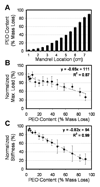
Figure 4. The composition and tensile properties of composite scaffolds can be tuned along the length of the mandrel
(A) Off-setting spinnerets results in a graded fiber sheet ranging from nominal (~5%) to ~90% PEO content along the mandrel as determined by PS mass loss. Scaffold % PEO content correlated with maximum stress (B) and modulus (C) when samples were tested in the fiber direction. Correlations were significant with p<0.001; n=3/group.
Figure 5
Figure 5. Increasing removal of sacrificial fiber content promotes mesenchymal stem cell (MSC) infiltration into composite fibrous scaffolds
Gross morphology (top row) and DAPI-stained cross-sections (bottom row) of MSC-seeded scaffolds with varying % PEO contents (% mass loss) after three weeks of in vitro culture. Scale bars: 5mm (top), 500µm (bottom).
Figure 6
Figure 6. Quantification of MSC infiltration into composite scaffolds as a function of sacrificial fiber content
Corresponding phase (A) and fluorescent (B) images of DAPI-stained construct cross-sections were used to evaluate cellular infiltration into composite scaffolds. (C) The average % infiltration increased in scaffolds above a threshold of ~40% PEO content in the as-spun scaffolds. To quantify cell distribution, infiltration distance was binned with respect to scaffold thickness, as shown in (B). (D) Comparisons of the lowest (~5%) and highest (~60%) PEO content constructs analyzed showed significantly higher fractions of cells within the more central regions of the scaffolds with increased PEO content. (E) The degree of cellular infiltration (as indicated by the % of total cells in each bin) showed a linear correlations with of % PEO content across a range of scaffold compositions. Diamond-ended bars (◆) indicate significance differences observed with p<0.05. A total of six samples were analyzed at each level of PEO content.
Figure 7
Figure 7. A simple model of composite scaffolds with increasing sacrifical fiber revmoval shows a reduction in pore number, but an increase in average pore size
(A) Example composite scaffold layers representing scaffolds with 10, 50, and 90% sacrificial PEO fibers. (B) Increasing the PEO fiber fraction decreases the total number of pores (●) while increasing the average pore area (◆). (C) Pore area distribution shifts towards higher pore sizes with increasing PEO content. Data were averaged from 20 model iterations, each with a randomly generated fiber population.

References

    1. Lynch HA, Johannessen W, Wu JP, Jawa A, Elliott DM. Effect of fiber orientation and strain rate on the nonlinear uniaxial tensile material properties of tendon. J Biomech Eng. 2003;125(5):726–731. - PubMed
    1. Setton LA, Guilak F, Hsu EW, Vail TP. Biomechanical factors in tissue engineered meniscal repair. Clin Orthop Relat Res. 1999;(367 Suppl):S254–S272. - PubMed
    1. Holzapfel GA, Schulze-Bauer CA, Feigl G, Regitnig P. Single lamellar mechanics of the human lumbar anulus fibrosus. Biomech Model Mechanobiol. 2005;3(3):125–140. - PubMed
    1. Newman AP, Anderson DR, Daniels AU, Dales MC. Mechanics of the healed meniscus in a canine model. Am J Sports Med. 1989;17(2):164–175. - PubMed
    1. Beredjiklian PK, Favata M, Cartmell JS, Flanagan CL, Crombleholme TM, Soslowsky LJ. Regenerative versus reparative healing in tendon: a study of biomechanical and histological properties in fetal sheep. Ann Biomed Eng. 2003;31(10):1143–1152. - PubMed

Publication types