Comparative Study
. 2008 Apr;149(1):12-9.
doi: 10.1016/j.jviromet.2008.01.016. Epub 2008 Mar 4.

Development and validation of a chemiluminescent immunodetection assay amenable to high throughput screening of antiviral drugs for Nipah and Hendra virus

Affiliations
Comparative Study

Development and validation of a chemiluminescent immunodetection assay amenable to high throughput screening of antiviral drugs for Nipah and Hendra virus

Mohamad Aljofan et al. J Virol Methods. 2008 Apr.

Abstract

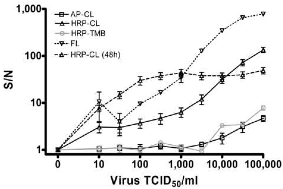
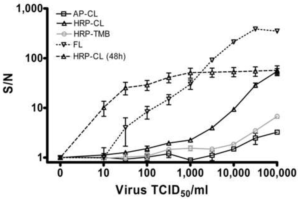
There are currently no antiviral drugs approved for the highly lethal Biosafety Level 4 pathogens Nipah and Hendra virus. A number of researchers are developing surrogate assays amenable to Biosafety Level 2 biocontainment but ultimately, the development of a high throughput screening method for directly quantifying these viruses in a Biosafety Level 4 environment will be critical for final evaluation of antiviral drugs identified in surrogate assays, in addition to reducing the time required for effective antiviral drug development. By adapting an existing immunoplaque assay and using enzyme linked immunodetection in a microtitre plate format, the current experiments describe a simple two step assay protocol involving an overnight virus inoculation of Vero cell monolayers (with or without antiviral drug treatment) at Biosafety Level 4, followed by cell fixation and virus inactivation enabling removal of plates from the Biosafety Level 4 laboratory and a subsequent immunodetection assay using a chemiluminescent horse radish peroxidase substrate to be performed at Biosafety Level 2. The analytical sensitivity (limit of detection) of this assay is 100 tissue culture infectious dose50/ml of either Nipah or Hendra virus. In addition this assay enables linear quantitation of virus over three orders of magnitude and is unaffected by dimethyl sulfoxide concentrations of 1% or less. Intra-assay coefficients of variation are acceptable (less than 20%) when detecting a minimum of 1000 tissue culture infectious dose50/ml of either virus although inter-assay variation is considerably greater. By an assessment of efficacies of the broad spectrum antiviral Ribavirin and an experimental fusion inhibitory peptide, this assay reveals a good correlation with previously published fluorescent immunodetection assays. The current experiments describe for the first time, a high throughput screening method amenable for direct assessment of live henipavirus antiviral drug activity.


Figures

Figure 1
Figure 1. Detection of NiV (a) and HeV (b) via immunodetection assays
½ log dilutions of virus (100ul) were incubated with 40,000 Vero cells per well for 24 hours at 37°C and 5% CO2. Monolayers were fixed with methanol, air dried and immunostained with anti-NiV-nucleoprotein polyclonal antisera (1:1000) followed by conjugated secondary antibodies (1:1000 – 1:2000). AP-CL, Alkaline Phosphatase conjugate with chemiluminescent substrate detection (n=4 replicate wells). HRP-CL (n=10), Horse Radish Peroxidase conjugate with chemiluminescent detection. HRP-TMB, Horse Radish Peroxidase conjugate with 3,3’,5,5’-Tetramethylbenzidine detection (A450nm, n=5). FL, Alexa-Fluor 488 conjugate (n=3). CL HRP (48h) as for HRP-CL after 48 hour virus infection (n=9). S/N, Signal:noise ratios calculated as signal/background values. Values are expressed as the Mean +/− S.E.
Figure 2
Figure 2. Effect of DMSO on NiV infection
½ log dilutions of virus were incubated as described in Figure 1 (24 hours) in the presence or absence of 1%, 2.5% and 5% DMSO (n=12 replicate wells). Cell monolayers were fixed, immunolabeled and detected using the HRP-CL assay. S/N, Signal:noise ratios. Values are expressed as the Mean +/− S.E.
Figure 3
Figure 3. Effect of plate storage on assay detection
½ log dilutions of NiV and HeV were incubated, immunolabeled and detected as described in Figure 1 (24 hours, HRP-CL). 4 plates containing 3 replicate wells were assayed immediately and compared to 4 plates stored at 4°C for 1 week prior to assay. Corresponding raw signal values (RLU, relative light units) for each virus dilution were plotted as Fresh (assayed immediately) vs Stored (assayed after storage). Correlation coefficients for each virus are indicated.
Figure 4
Figure 4. Effect of virus concentration on assay coefficient of variation
½ log dilutions of NiV and HeV were incubated, immunolabeled and detected as described in Figure 1 (24 hours, HRP-CL). Coefficient of variation is plotted against virus concentration. Intra assay values are averages of 3 plates containing 6 replicates. Inter assay values are averages of 6 replicates. Values are expressed as the Mean +/− S.E.
Figure 5
Figure 5. Evaluation of Henipavirus inhibitors using HRP-chemiluminescent (a) and fluorescent (b) detection
NiV (m.o.i.= 0.25) was inoculated onto Vero cells preincubated with log dilutions of HPIV3 F protein derived peptide (Wt), Scrambled HPIV3 F peptide (Sc) or Ribavirin (Rib), incubated for 24 hours at 37°C and 5% CO2 then fixed, immunolabeled and detected as described in Figure 1 (HRP-CL). % inhibition was determined compared to untreated, infected wells and values are expressed as the Mean +/− S.E (n=8 replicate wells). Non-linear regression analysis was performed (GraphPad Prism) to determine the 50% inhibitory concentration (IC50).

References

    1. Atamna H, Boyle K. Amyloid-beta peptide binds with heme to form a peroxidase: relationship to the cytopathologies of Alzheimer's disease. Proc Natl Acad Sci U S A. 2006;103:3381–3386. - PMC - PubMed
    1. Bellini WJ, Harcourt BH, Bowden N, Rota PA. Nipah virus: an emergent paramyxovirus causing severe encephalitis in humans. J Neurovirol. 2005;11:481–487. - PubMed
    1. Chong H, Kamarulzaman A, Tan C, Goh K, Thayaparan T, Kunjapan SR, Chew N, Chua K, Lam S. Treatment of Acute Nipah Encephalitis With Ribavirin. Annals of Neurology. 2001;49:810–813. - PubMed
    1. Chua KB. Nipah virus outbreak in Malaysia. J Clin Virol. 2003;26:265–275. - PubMed
    1. Chua KB, Goh KJ, Wong KT, Kamarulzaman A, Tan PS, Ksiazek TG, Zaki SR, Paul G, Lam SK, Tan CT. Fatal encephalitis due to Nipah virus among pig-farmers in Malaysia. Lancet. 1999;354:1257–1259. - PubMed

Publication types

MeSH terms

Substances

LinkOut - more resources