. 2008 Mar 27;152(3):773-84.
doi: 10.1016/j.neuroscience.2008.01.007. Epub 2008 Jan 12.

Tolerance to 3,4-methylenedioxymethamphetamine in rats exposed to single high-dose binges

Affiliations

Tolerance to 3,4-methylenedioxymethamphetamine in rats exposed to single high-dose binges

M H Baumann et al. Neuroscience. .

Abstract

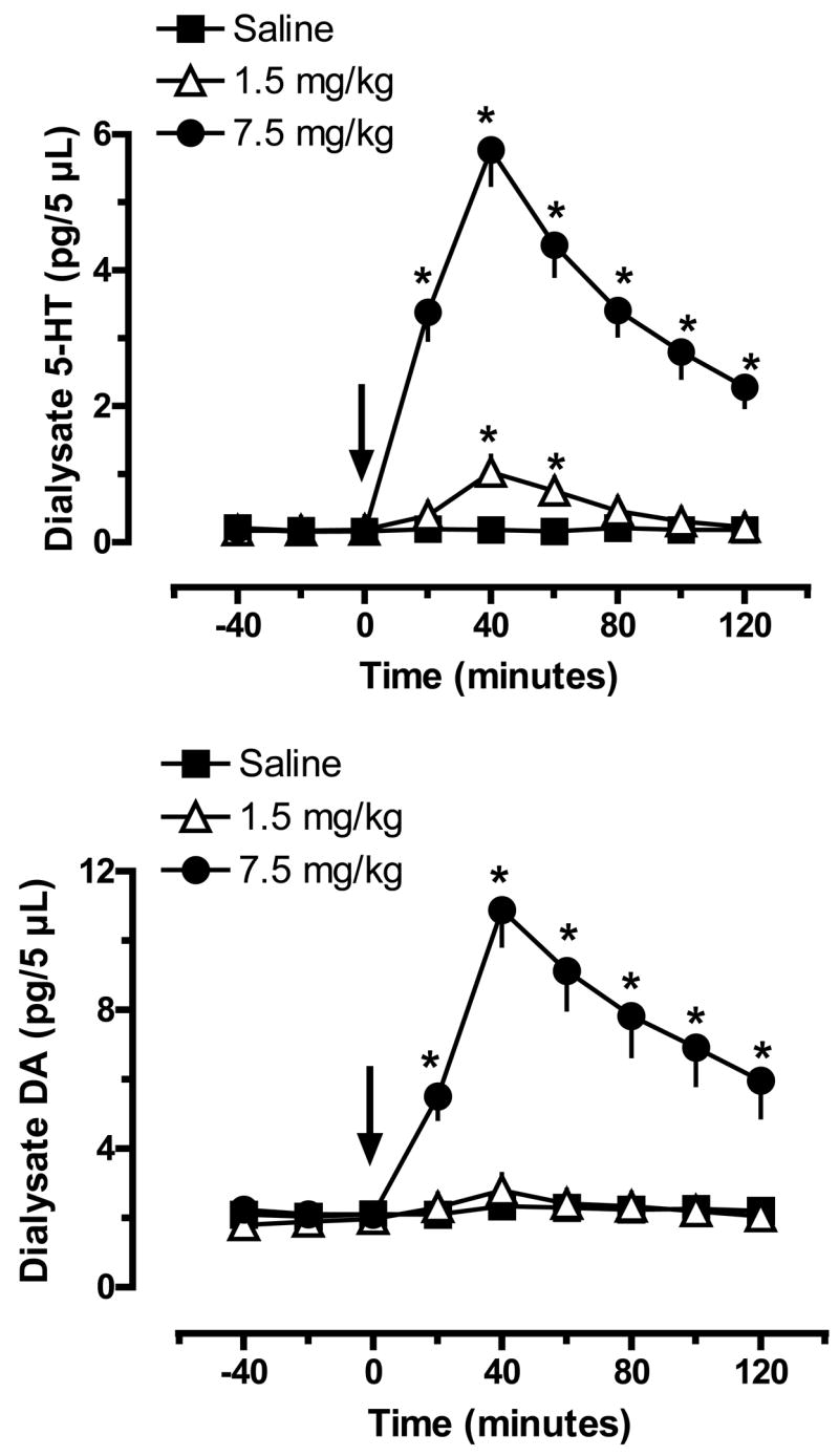
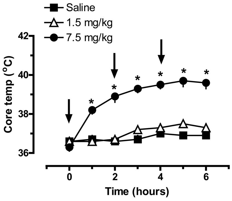
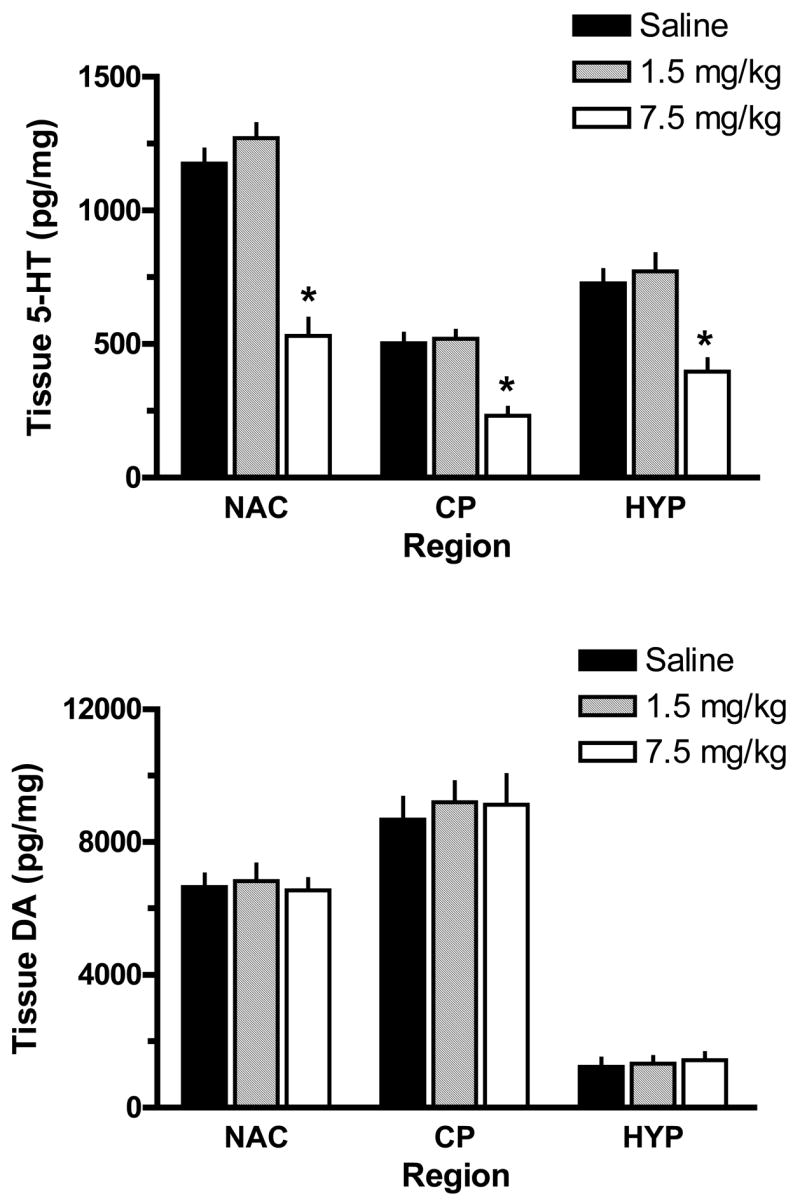
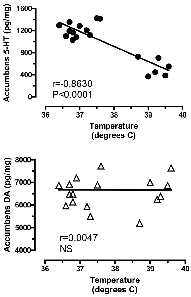
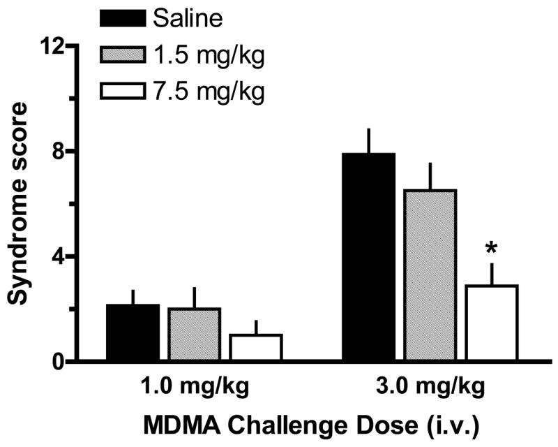
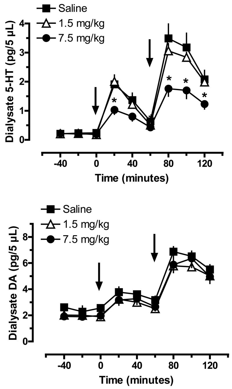
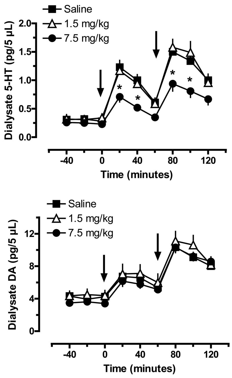
3,4-Methylenedioxymethamphetamine (MDMA or ecstasy) stimulates the transporter-mediated release of monoamines, including 5-HT. High-dose exposure to MDMA causes persistent 5-HT deficits (e.g. depletion of brain 5-HT) in animals, yet the functional and clinical relevance of such deficits are poorly defined. Here we examine functional consequences of MDMA-induced 5-HT depletions in rats. Male rats received binges of three i.p. injections of MDMA or saline, one injection every 2 h; MDMA was given at a threshold pharmacological dose (1.5 mg/kgx3, low dose) or at a fivefold higher amount (7.5 mg/kgx3, high dose). One week later, jugular catheters and intracerebral guide cannulae were implanted. Two weeks after binges, rats received acute i.v. challenge injections of 1 and 3 mg/kg MDMA. Neuroendocrine effects evoked by i.v. MDMA (prolactin and corticosterone secretion) were assessed via serial blood sampling, while neurochemical effects (5-HT and dopamine release) were assessed via microdialysis in brain. MDMA binges elevated core temperatures only in the high-dose group, with these same rats exhibiting approximately 50% loss of forebrain 5-HT 2 weeks later. Prior exposure to MDMA did not alter baseline plasma hormones or dialysate monoamines, and effects of i.v. MDMA were similar in saline and low-dose groups. By contrast, rats pretreated with high-dose MDMA displayed significant reductions in evoked hormone secretion and 5-HT release when challenged with i.v. MDMA. As tolerance developed only in rats exposed to high-dose binges, hyperthermia and 5-HT depletion are implicated in this phenomenon. Our results suggest that MDMA tolerance in humans may reflect 5-HT deficits which could contribute to further dose escalation.


Figures

Figure 1
Figure 1
Effects of MDMA administration on extracellular levels of 5-HT (top panel) and DA (bottom panel) in rat n. accumbens. Male rats undergoing in vivo microdialysis received single ip injections of saline, 1.5 mg/kg or 7.5 mg/kg MDMA at time zero. Dialysate samples were collected at 20 min intervals and assayed for 5-HT and DA by HPLC-ECD. Data are pg/5 μL sample expressed as mean±SEM for N=6 rats/group. * = P < 0.05 compared to saline control at a given time point.
Figure 2
Figure 2
Effects of MDMA binge administration on body temperature in rats. Male rats received 3 ip injections of saline, 1.5 mg/kg (low dose) or 7.5 mg/kg MDMA (high dose); injections were given at time zero, 2h and 4h. Core body temperature was monitored hourly by insertion of a temperature probe into the colon. Data are degrees Celsius expressed as mean±SEM for N=6 rats/group. * = P < 0.05 compared to saline control at a given time point.
Figure 3
Figure 3
Effects of MDMA binge administration on tissue levels of 5-HT (top panel) and DA (bottom panel) in microdissected rat brain regions. Male rats received 3 ip injections of saline, 1.5 mg/kg (low dose) or 7.5 mg/kg MDMA (high dose). Rats were euthanized 2 weeks later, and tissue was dissected from n. accumbens (NAC), caudate-putamen (CP) and mediobasal hypothalalmus (HYP). Concentrations of 5-HT and DA were quantified by HPLC-ECD. Data are pg/mg wet weight expressed as mean±SEM for N= 6 rats/group. * = P < 0.05 compared to saline control for a specified brain region.
Figure 4
Figure 4
Correlations between acute body temperature and post-mortem tissue levels of 5-HT (top panel) and DA (bottom panel) in rat n. accumbens measured two weeks later. Body temperatures represent average values obtained from 1–6 h after MDMA or saline injections, as shown in Fig. 2. Post-mortem tissue levels of 5-HT and DA were measured two weeks later by HPLC-ECD, as shown in Fig. 3. Data points represent average temperature vs. tissue amine concentration from individual rats (N=18 rats), and Pearson correlation coefficients (r) are given.
Figure 5
Figure 5
Plasma concentrations of prolactin (top panel) and corticosterone (bottom panel) in rats pretreated with saline or MDMA binges. Male rats received 3 ip injections of saline, 1.5 mg/kg (low dose) or 7.5 mg/kg MDMA (high dose). Two weeks later, rats were given iv challenge injections of 1 mg/kg MDMA at time zero, followed by 3 mg/kg MDMA at 60 min. Blood samples were withdrawn immediately before and at 15, 30, 60, 75, 90 and 120 min after the first iv injection. Plasma hormones were assayed by double-antibody RIA methods. Data are ng/ml expressed as mean±SEM for N=8 rats/group. * = P < 0.05 compared to saline-pretreated control at a given time point.
Figure 6
Figure 6
5-HT behavioral syndrome in rats pretreated with saline or MDMA binges. Male rats received 3 ip injections of saline, 1.5 mg/kg (low dose) or 7.5 mg/kg MDMA (high dose). Two weeks later, rats were given iv challenge injections of 1 mg/kg MDMA at time zero, followed by 3 mg/kg MDMA at 60 min. The occurrence of flat-body posture and forepaw treading were scored after each dose of acute iv MDMA. Data are syndrome scores expressed as mean±SEM for N=8 rats/group. * = P < 0.05 compared to saline-pretreated control at the corresponding challenge dose of MDMA.
Figure 7
Figure 7
Extracellular levels of 5-HT (top panel) and DA (bottom panel) in n. accumbens of rats pretreated with saline or MDMA binges. Male rats received 3 ip injections of saline, 1.5 mg/kg (low dose) or 7.5 mg/kg MDMA (high dose). Two weeks later, rats undergoing in vivo microdialysis were given iv challenge injections of 1 mg/kg MDMA at time zero, followed by 3 mg/kg MDMA at 60 min. Dialysate samples collected at 20 min intervals were assayed for 5-HT and DA by microbore HPLC-ECD. Data are pg/5 μL expressed as mean±SEM for N=8 rats/group. * = P < 0.05 compared to saline-pretreated control at a given time point.
Figure 8
Figure 8
Extracellular levels of 5-HT (top panel) and DA (bottom panel) in caudate n. of rats pretreated with saline or MDMA binges. Male rats received 3 ip injections of saline, 1.5 mg/kg (low dose) or 7.5 mg/kg MDMA (high dose). Two weeks later, rats undergoing in vivo microdialysis were given iv challenge injections of 1 mg/kg MDMA at time zero, followed by 3 mg/kg MDMA at 60 min. Dialysate samples collected at 20 min intervals were assayed for 5-HT and DA by microbore HPLC-ECD. Data are pg/5 μL expressed as mean±SEM for N=8 rats/group. * = P < 0.05 compared to saline-pretreated control at a given time point.

References

    1. Ball KT, Budreau D, Rebec GV. Acute effects of 3,4-methylenedioxymethamphetamine on striatal single-unit activity and behavior in freely moving rats: differential involvement of dopamine D(1) and D(2) receptors. Brain Res. 2003;994:203–215. - PubMed
    1. Bankson MG, Cunningham KA. 3,4-Methylenedioxymethamphetamine (MDMA) as a unique model of serotonin receptor function and serotonin-dopamine interactions. J Pharmacol Exp Ther. 2001;297:846–852. - PubMed
    1. Battaglia G, Yeh SY, O’Hearn E, Molliver ME, Kuhar MJ, De Souza EB. 3,4-Methylenedioxymethamphetamine and 3,4-methylenedioxyamphetamine destroy serotonin terminals in rat brain: quantification of neurodegeneration by measurement of [3H]paroxetine-labeled serotonin uptake sites. J Pharmacol Exp Ther. 1987;242:911–916. - PubMed
    1. Baumann MH, Ayestas MA, Rothman RB. Functional consequences of central serotonin depletion produced by repeated fenfluramine administration in rats. J Neurosci. 1998;18:9069–9077. - PMC - PubMed
    1. Baumann MH, Clark RD, Budzynski AG, Partilla JS, Blough BE, Rothman RB. N-substituted piperazines abused by humans mimic the molecular mechanism of 3,4-methylenedioxymethamphetamine (MDMA, or ‘Ecstasy’) Neuropsychopharmacology. 2005;30:550–560. - PubMed

Publication types

MeSH terms