. 2008 Apr 1;177(3):188-97.
doi: 10.1016/j.toxlet.2008.01.010. Epub 2008 Jan 31.

Mitochondrial protein thiol modifications in acetaminophen hepatotoxicity: effect on HMG-CoA synthase

Affiliations

Mitochondrial protein thiol modifications in acetaminophen hepatotoxicity: effect on HMG-CoA synthase

Kelly K Andringa et al. Toxicol Lett. .

Abstract

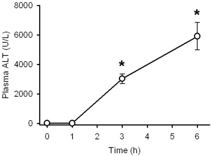
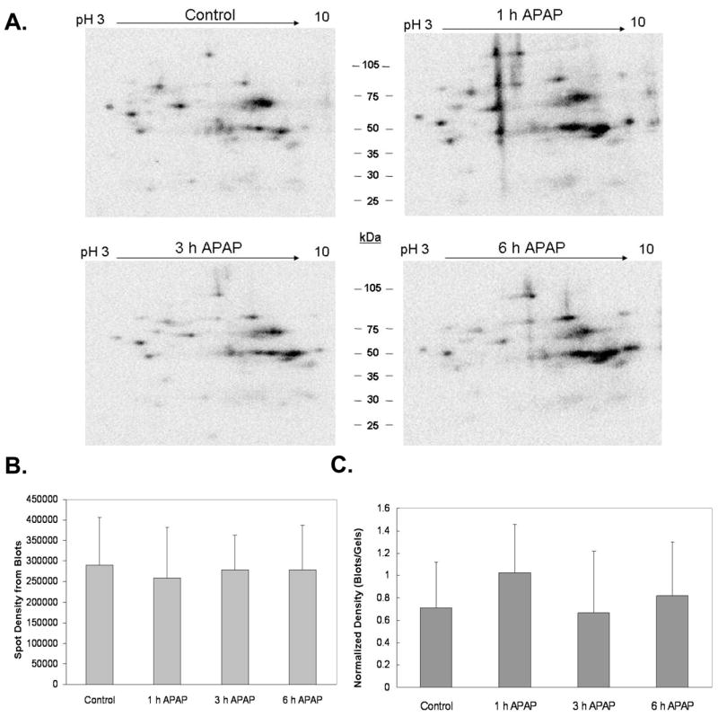
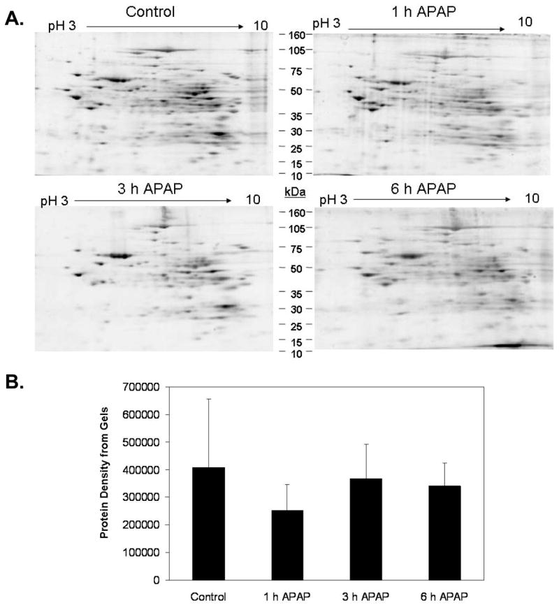
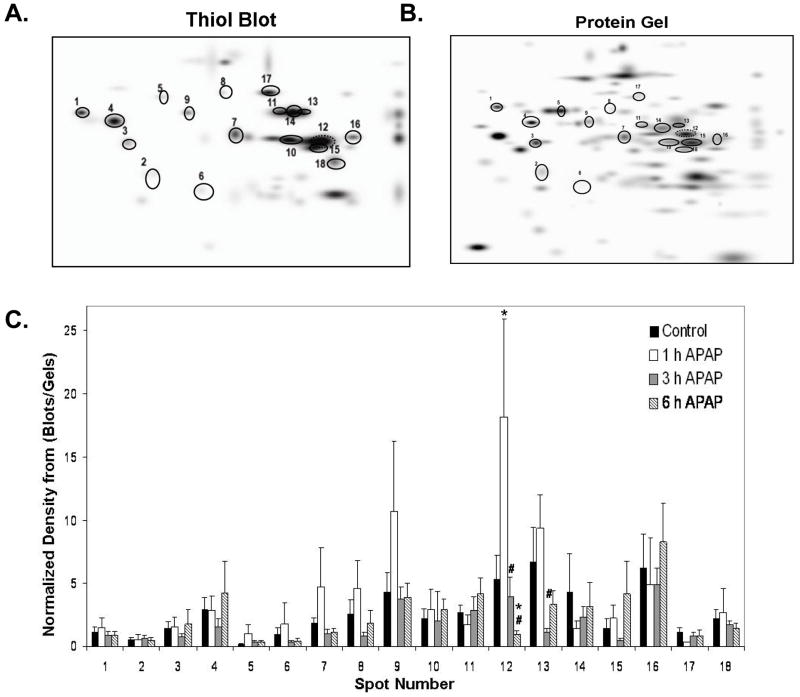
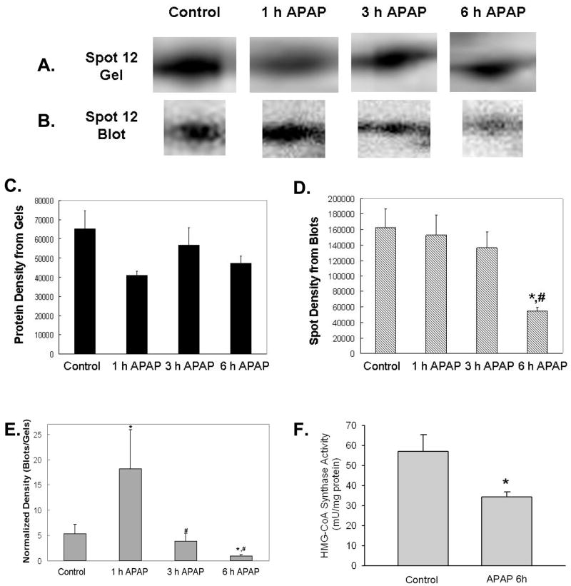
Acetaminophen (APAP) overdose is the leading cause of drug related liver failure in many countries. N-acetyl-p-benzoquinone imine (NAPQI) is a reactive metabolite that is formed by the metabolism of APAP. NAPQI preferentially binds to glutathione and then cellular proteins. NAPQI binding is considered an upstream event in the pathophysiology, especially when binding to mitochondrial proteins and therefore leads to mitochondrial toxicity. APAP caused a significant increase in liver toxicity 3h post-APAP administration as measured by increased serum alanine aminotransferase (ALT) levels. Using high-resolution mitochondrial proteomics techniques to measure thiol and protein changes, no significant change in global thiol levels was observed. However, 3-hydroxy-3-methylglutaryl coenzyme A synthase 2 (HMG-CoA synthase) had significantly decreased levels of reduced thiols and activity after APAP treatment. HMG-CoA synthase is a key regulatory enzyme in ketogenesis and possesses a number of critical cysteines in the active site. Similarly, catalase, a key enzyme in hydrogen peroxide metabolism, also showed modification in protein thiol content. These data indicate post-translational modifications of a few selected proteins involved in mitochondrial and cellular regulation of metabolism during liver toxicity after APAP overdose. The pathophysiological relevance of these limited changes in protein thiols remains to be investigated.


Figures

Figure 1
Figure 1. Increased serum ALT levels in response to Acetaminophen (APAP) exposure
APAP treatment (300 mg/kg) causes liver injury as indicated by significant increases in plasma ALT levels at 3 and 6 h after treatment. Data represent mean ± SE of n = 5 animals per time point. *p < 0.05 (compared to time zero)
Figure 2
Figure 2. Effect of APAP treatment on mitochondrial protein thiol content
Mitochondria isolated from liver of mice treated with 300 mg/kg APAP were incubated with BIAM, a compound that labels reduced thiols. Labeled proteins were separated by 2D IEF/SDS-PAGE and gels were subjected to immunoblotting to visualize BIAM-labeled proteins. Immunoblot analysis showed no significant difference in thiol labeling intensity between control and APAP treated groups over the time period of the experiment. Panel A shows representative BIAM blots for each treatment group, panel B shows the density of the BIAM blots, and panel C shows the normalized density of total BIAM labeled protein from blots as compared to the total protein density of matched gels shown in Figure 3. Data represent mean ± SD of n = 3–5 animals per time point.
Figure 3
Figure 3. Effect of APAP treatment on mitochondrial proteins
Mitochondria isolated from liver of mice treated with 300 mg/kg APAP were subjected to 2D IEF/SDS-PAGE proteomics protocol. Gels were stained with Sypro Ruby protein stain. Panel A shows representative gels for each treatment. Panel B, Image analysis revealed that mitochondrial protein content in APAP treated groups was no different when compared to the untreated control. Data represent mean ± SD of n = 3–5 animals per time point.
Figure 4
Figure 4. Master maps of BIAM-labeled proteins and matched proteins from 2D gels
The spots circled on these master images represent BIAM-labeled protein spots (A) that were matched across all treatment groups. The corresponding proteins for these BIAM-labeled proteins on gels are shown in Panel B. Densitometry of individual BIAM-labeled protein thiols from blots and protein gels were calculated and normalized values of BIAM-labeled proteins is shown in Panel C (blot spots/gel spots). Two proteins showed significant differences in thiol labeling as a consequence of APAP treatment. Data represent mean ± SD of n = 3–5 animals per time point. *p < 0.05, compared to control; #p < 0.05, compared to 1 h APAP treatment.
Figure 5
Figure 5. Decreased thiol content and activity in HMG-CoA synthase in response to APAP treatment
Mitochondrial samples isolated from the liver of mice treated with 300 mg/kg APAP were analyzed as described in Figures 2 and 3. APAP treatment resulted in significant changes in the thiol content of HMG-CoA synthase. Panel A shows representative HMG-CoA synthase protein from 2D gels with corresponding BIAM-labeling shown in Panel B for each treatment. Panels C and D show the densitometry of HMG-CoA synthase protein and thiol content, respectively. Panel E shows the normalized blot density over protein density. There was a significant increase in thiol content at 1 h with significant decreases in thiol content of HMG-CoA synthase at both 3 and 6 h time points. Panel F shows the decrease in HMG-CoA synthase activity at 6 h time point. HMG-CoA synthase activity was determined as described in the methods sections. Data (panel A–E) represent mean ± SD of n = 3–5 animals per time point. Data (panel F) represent mean ± SE of n = 6 animals per group. *p < 0.05, compared to control; #p < 0.05, compared to 1 h APAP treatment.

References

    1. Bailey SM, Landar A, Darley-Usmar V. Mitochondrial proteomics in free radical research. Free Radic Biol Med. 2005;38:175–188. - PubMed
    1. Bajt ML, Knight TR, Farhood A, Jaeschke H. Scavenging peroxynitrite with glutathione promotes regeneration and enhances survival during acetaminophen-induced liver injury in mice. J Pharmacol Exper Therap. 2003;307:67–73. - PubMed
    1. Bajt ML, Cover C, Lemasters JJ, Jaeschke H. Nuclear translocation of endonuclease G and apoptosis-inducing factor during acetaminophen-induced liver cell injury. Toxicol Sci. 2006;94:217–225. - PubMed
    1. Barford D. The role of cysteine residues as redox-sensitive regulatory switches. Curr Opin Struct Biol. 2004;14:679–686. - PubMed
    1. Beer SM, Taylor ER, Brown SE, Dahm CC, Costa NJ, Runswick MJ, Murphy MP. Glutaredoxin 2 catalyzes the reversible oxidation and glutathionylation of mitochondrial membrane thiol proteins. J Biol Chem. 2004;279:47939–47951. - PubMed

Publication types

LinkOut - more resources