Comment
. 2008 Mar;5(3):427-35.
doi: 10.1016/j.hrthm.2007.12.019. Epub 2008 Feb 4.

Gain of function in IKs secondary to a mutation in KCNE5 associated with atrial fibrillation

Affiliations
Comment

Gain of function in IKs secondary to a mutation in KCNE5 associated with atrial fibrillation

Lasse S Ravn et al. Heart Rhythm. 2008 Mar.

Abstract

Background: Atrial fibrillation (AF) is the most common clinical arrhythmia and a major cause of cardiovascular morbidity and mortality. Among the gene defects previously associated with AF is a gain of function of the slowly activating delayed rectifier potassium current IKs, secondary to mutations in KCNQ1. Coexpression of KCNE5, the gene encoding the MiRP4 beta-subunit, has been shown to reduce IKs.

Objective: The purpose of this study was to test the hypothesis that mutations in KCNE5 are associated with AF in a large cohort of patients with AF.

Methods: One-hundred fifty-eight patients with AF were screened for mutations in the coding region of KCNE5.

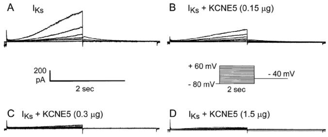
Results: A missense mutation involving substitution of a phenylalanine for leucine at position 65 (L65F) was identified in one patient. This patient did not have a history of familial AF, and neither KCNQ1 nor KCNE2 mutations were found. Transient transfection of Chinese hamster ovary (CHO) cells expressing IKs(KCNQ1+KCNE1) with KCNE5 suppressed the developing and tail currents of IKs in a concentration-dependent manner. Transient transfection with KCNE5-L65F failed to suppress IKs, yielding a current indistinguishable from that recorded in the absence of KCNE5. Developing currents recorded during a test pulse to +60 mV and tail currents recorded upon repolarization to -40 mV both showed a significant concentration-dependent gain of function in IKs with expression of KCNE5-L65F vs KCNE5-WT.

Conclusion: The results of this study suggest that a missense mutation in KCNE5 may be associated with nonfamilial or acquired forms of AF. The arrhythmogenic mechanism most likely is a gain of function of IKs.


Figures

Figure 1
Figure 1
Mutation screening of KCNE5. A: DNA sequence chromatogram showing the wild type (WT) and the corresponding amino acid sequence. B: DNA sequence chromatogram showing the heterozygous mutation (in bold) and the corresponding amino acid sequence. A heterozygous substitution of T for C in nucleotide 193 predicts substitution of phenylalanine (TTC) for leucine (CTC) in codon 65 (L65F). C: ECG of the proband recorded during normal sinus rhythm between episodes of atrial fibrillation.
Figure 2
Figure 2
Representative current trace recorded from IKs/CHO cells transiently transfected with KCNE5-WT. A: Current from stable cell line of IKs/CHO. B: Current from IKs/CHO transfected with 0.15 μg KCNE5-WT. C: Current from IKs/CHO transfected with 0.3 μg KCNE5-WT. D: Current from IKs/CHO transfected with 1.5 μg KCNE5-WT. Pulse protocol is shown in the inset in the center right.
Figure 3
Figure 3
Current-voltage relationship of developing and tail currents in the absence and presence of 0.15 to 1.5 μg KCNE5-WT. A: Current-voltage relationship of peak developing current. B: Current-voltage relationship of tail current measured upon repolarization to -40 mV. C: Bar graphs showing current densities of peak developing current measured during test pulse to +60 mV. D: Bar graphs showing current densities of tail current measured upon repolarization to -40 mV following a test pulse to +60 mV. *P <.05; **P <.01 vs IKs.
Figure 4
Figure 4
Current-voltage relationship of developing and tail currents in the absence and presence of 1.5 μg KCNE5-WT or KCNE5-L65F. A: Representative current trace of IKs/CHO cells transfected with 1.5 μg WT KCNE5. B: Representative current trace of IKs/CHO cells transfected with 1.5 μg L65F KCNE5. Pulse protocol is shown in the inset in the center right. C: Current-voltage relationship of peak developing current. D: Current-voltage relationship of tail current measured upon repolarization to -40 mV. *P <.05 vs IKs+KCNE5-WT.
Figure 5
Figure 5
Effect of 0.3 and 1.5 μg KCNE5-WT or KCNE5-L65F on IKs. A: Bar graph showing density of peak developing current recorded during a test pulse to +60 mV. B: Bar graph showing density of tail currents recorded during repolarization to -40 mV following a test pulse to +60 mV. Statistical significance determined using analysis of variance. *P <.05; **P <.01.
Figure 6
Figure 6
Current-voltage relationship of developing and tail currents in cells cotransfected with equal amounts of KCNE5-WT and KCNE5-L65F. A: Representative current trace of IKs/CHO cells transfected with 0.75 μg WT KCNE5 and 0.75 μg L65F KCNE5. B: Representative current trace of IKs/CHO cells transfected with 1.5 μg WT KCNE5 and 1.5 μg L65F KCNE5. C: Current-voltage relationship of peak developing current. D: Current-voltage relationship of tail current measured upon repolarization to -40 mV. E: Bar graph showing density of peak developing current recorded during a test pulse to +60 mV (IKs; IKs+1.5 μg KCNE5-WT; IKs+0.75 μg KCNE5-WT+0.75 μg KCNE5-L65F; IKs+1.5 μg KCNE5-WT+1.5 μg KCNE5-L65F). F: Bar graph showing density of tail currents recorded during repolarization to -40 mV following a test pulse to +60 mV (IKs; IKs+1.5 μg KCNE5-WT; IKs+0.75 μg KCNE5-WT+0.75 μg KCNE5-L65F; IKs+1.5 μg KCNE5-WT+1.5 μg KCNE5-L65F).

Comment on

  • Ion channel mutations in AF: signal or noise?
    Ellinor PT, MacRae CA. Ellinor PT, et al. Heart Rhythm. 2008 Mar;5(3):436-7. doi: 10.1016/j.hrthm.2008.01.014. Epub 2008 Jan 17. Heart Rhythm. 2008. PMID: 18313603 Free PMC article. No abstract available.

References

    1. Frost L, Engholm G, Moller H, et al. Decrease in mortality in patients with a hospital diagnosis of atrial fibrillation in Denmark during the period 1980-1993. Eur Heart J. 1999;20:1592–1599. - PubMed
    1. Friberg J, Scharling H, Gadsboll N, et al. Sex-specific increase in the prevalence of atrial fibrillation (The Copenhagen City Heart Study) Am J Cardiol. 2003;92:1419–1423. - PubMed
    1. Frost L, Engholm G, Johnsen S, et al. Incident stroke after discharge from the hospital with a diagnosis of atrial fibrillation. Am J Med. 2000;108:36–40. - PubMed
    1. Grogan M, Smith HC, Gersh BJ, et al. Left ventricular dysfunction due to atrial fibrillation in patients initially believed to have idiopathic dilated cardiomyopathy. Am J Cardiol. 1992;69:1570–1573. - PubMed
    1. Darbar D, Herron KJ, Ballew JD, et al. Familial atrial fibrillation is a genetically heterogeneous disorder. J Am Coll Cardiol. 2003;41:2185–2192. - PubMed

Publication types

Substances