

Review
doi: 10.1016/j.hfc.2007.10.007.
Molecular basis of diastolic dysfunction
Affiliations
- PMID: 18313621
- PMCID: PMC2705955
- DOI: 10.1016/j.hfc.2007.10.007
Item in Clipboard
Review
Molecular basis of diastolic dysfunction
Heart Fail Clin.
2008 Jan.
Abstract
Diastolic dysfunction is characterized by prolonged relaxation, increased filling pressure, decreased contraction velocity, and reduced cardiac output. Phenotypical features of diastolic dysfunction can be observed at the level of the isolated myocyte. This article reviews the cellular mechanisms that control relaxation at the level of the myocyte in the healthy situation and discusses the alterations that can affect physiologic function during disease. It focuses specifically on the mechanisms that regulate intracellular calcium handling, and the response of the myofilaments to calcium, including the changes in these components that can contribute to diastolic dysfunction.
Figures
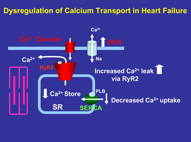
Potential contributors to Ca2+ dysregulation in failing heart muscle. A decreased expression and activity of SERCA pump could explain slowed rate of Ca2+ uptake and prolongation of muscle relaxation. Enhanced NCX expression and activity could increase Ca2+ efflux and compete with SERCA pump, reducing SR Ca2+ stores. RyR2 phosphorylation by PKA or CaMKII causes increased opening and Ca2+ leak, further contributing to loss of Ca2+. A net loss of Ca2+ could contribute to both systolic and diastolic dysfunction.
References
-
- Borlaug BA, Kass DA. Mechanisms of diastolic dysfunction in heart failure. Trends Cardiovasc Med. 2006 Nov;16(8):273–279. - PubMed
-
- Kass DA, Bronzwaer JG, Paulus WJ. What mechanisms underlie diastolic dysfunction in heart failure? Circ Res. 2004 Jun 25;94(12):1533–1542. - PubMed
-
- Cooper Gt. Cytoskeletal networks and the regulation of cardiac contractility: microtubules, hypertrophy, and cardiac dysfunction. Am J Physiol Heart Circ Physiol. 2006 Sep;291(3):H1003–1014. - PubMed
-
- Zile MR, Baicu CF, Bonnema DD. Diastolic heart failure: definitions and terminology. Prog Cardiovasc Dis. 2005 Mar–Apr;47(5):307–313. - PubMed
-
- Fabiato A, Fabiato F. Calcium and cardiac excitation-contraction coupling. Annu Rev Physiol. 1979;41:473–484. - PubMed
Publication types
MeSH terms
Substances
Grants and funding
LinkOut - more resources
Full Text Sources
Other Literature Sources
Medical
