

Analysis of apoptosis by cytometry using TUNEL assay
- PMID: 18314056
- PMCID: PMC2295206
- DOI: 10.1016/j.ymeth.2007.11.008
Analysis of apoptosis by cytometry using TUNEL assay
Abstract
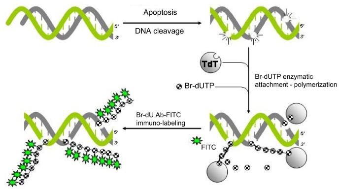
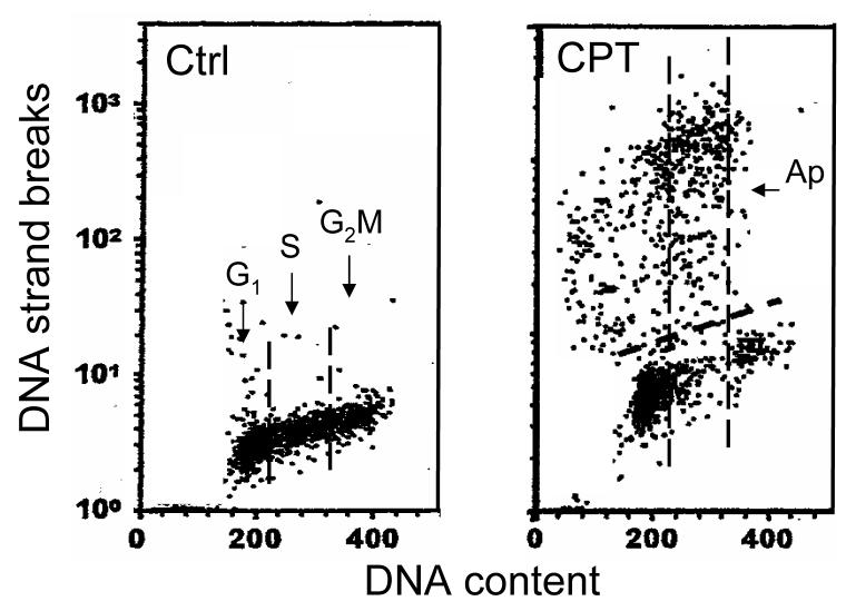
Activation of endonucleases that cleave chromosomal DNA preferentially at internucleosomal sections is a hallmark of apoptosis. DNA fragmentation revealed by the presence of a multitude of DNA strand breaks, therefore, is considered to be the gold standard for identification apoptotic cells. Several variants of the methodology that is based on fluorochrome-labeling of 3'-OH termini of DNA strand breaks in situ with the use of exogenous terminal deoxynucleotidyl transferase (TdT), commonly defined as the TUNEL assay, have been developed by us. This Chapter describes the variant based on strand breaks labeling with Br-dUTP that is subsequently detected immunocytochemically with Br-dUAb. Compared with other TUNEL variants the Br-dU-labeling assay offers the greatest sensitivity in detecting DNA breaks. Described also are modifications of the protocol that allow one to use other than Br-dUTP fluorochrome-tagged deoxynucleotides to label DNA breaks. Concurrent staining of DNA with propidium or 4',6-diamidino-2-phenylindole (DAPI) and multiparameter analysis of cells by flow- or laser scanning cytometry enables one to correlate induction of apoptosis with the cell cycle phase.
Figures
Similar articles
-
Detection of DNA strand breaks in apoptotic cells by flow- and image-cytometry.Methods Mol Biol. 2011;682:91-101. doi: 10.1007/978-1-60327-409-8_8. Methods Mol Biol. 2011. PMID: 21057923 Free PMC article.
-
Rapid Detection of DNA Strand Breaks in Apoptotic Cells by Flow- and Image-Cytometry.Methods Mol Biol. 2017;1644:139-149. doi: 10.1007/978-1-4939-7187-9_12. Methods Mol Biol. 2017. PMID: 28710760
-
Detection of end-stage apoptosis by ApopTag® TUNEL technique.Methods Mol Biol. 2015;1219:43-56. doi: 10.1007/978-1-4939-1661-0_5. Methods Mol Biol. 2015. PMID: 25308261
-
An overview of apoptosis assays detecting DNA fragmentation.Mol Biol Rep. 2018 Oct;45(5):1469-1478. doi: 10.1007/s11033-018-4258-9. Epub 2018 Jul 18. Mol Biol Rep. 2018. PMID: 30022463 Review.
-
Do TUNEL and Other Apoptosis Assays Detect Cell Death in Preclinical Studies?Int J Mol Sci. 2020 Nov 29;21(23):9090. doi: 10.3390/ijms21239090. Int J Mol Sci. 2020. PMID: 33260475 Free PMC article. Review.
Cited by
-
PNT2258, a novel deoxyribonucleic acid inhibitor, induces cell cycle arrest and apoptosis via a distinct mechanism of action: a new class of drug for non-Hodgkin's lymphoma.Oncotarget. 2016 Jul 5;7(27):42374-42384. doi: 10.18632/oncotarget.9872. Oncotarget. 2016. PMID: 27283896 Free PMC article.
-
Flavonol isolated from ethanolic leaf extract of Thuja occidentalis arrests the cell cycle at G2-M and induces ROS-independent apoptosis in A549 cells, targeting nuclear DNA.Cell Prolif. 2014 Feb;47(1):56-71. doi: 10.1111/cpr.12079. Epub 2013 Nov 23. Cell Prolif. 2014. PMID: 24267912 Free PMC article.
-
Curcumin-loaded γ-cyclodextrin liposomal nanoparticles as delivery vehicles for osteosarcoma.Nanomedicine. 2012 May;8(4):440-51. doi: 10.1016/j.nano.2011.07.011. Epub 2011 Aug 10. Nanomedicine. 2012. PMID: 21839055 Free PMC article.
-
Membrane protected apoptotic trophoblast microparticles contain nucleic acids: relevance to preeclampsia.Am J Pathol. 2008 Dec;173(6):1595-608. doi: 10.2353/ajpath.2008.080414. Epub 2008 Oct 30. Am J Pathol. 2008. PMID: 18974299 Free PMC article.
-
MicroRNA miR-497 is closely associated with poor prognosis in patients with cerebral ischemic stroke.Bioengineered. 2021 Dec;12(1):2851-2862. doi: 10.1080/21655979.2021.1940073. Bioengineered. 2021. PMID: 34152256 Free PMC article.
References
-
- Nagata S. Exp. Cell Res. 2000;256:12–18. - PubMed
-
- Enari M, Sakahira H, Yokoyama H, Okawa K, Iwamatsu A, Nagata S. Nature. 1998;391:943–50. S. - PubMed
-
- Kajstura M, Halicka HD, Pryjma J, Darzynkiewicz Z. Cytometry A. 2007;71A:125–131. - PubMed
-
- Gorczyca W, Bruno S, Darzynkiewicz RJ, Gong J, Darzynkiewicz Z. Int. J. Oncol. 1992;1:639–648. - PubMed
-
- Darzynkiewicz Z, Juan G, Li X, Gorczyca W, Murakami T, Traganos F. Cytometry. 1997;27:1–20. F. - PubMed
Publication types
MeSH terms
Substances
Grants and funding
LinkOut - more resources
Full Text Sources
Other Literature Sources
