. 2008 Apr;14(4):736-48.
doi: 10.1261/rna.824308. Epub 2008 Feb 26.

Role of SLV in SLI substrate recognition by the Neurospora VS ribozyme

Affiliations

Role of SLV in SLI substrate recognition by the Neurospora VS ribozyme

Patricia Bouchard et al. RNA. 2008 Apr.

Abstract

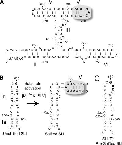
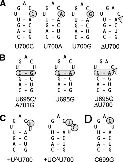
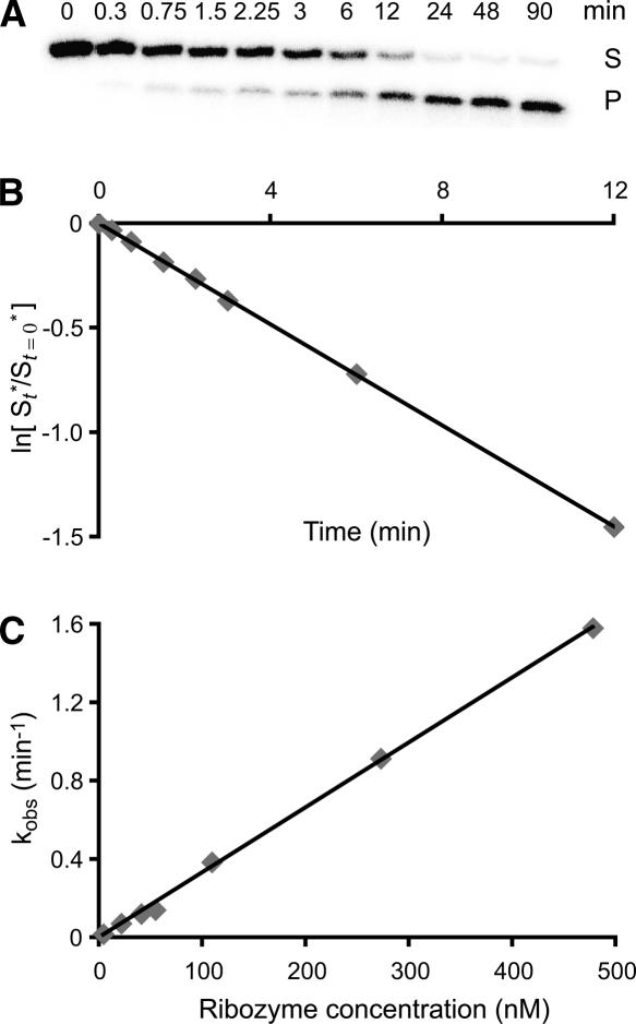
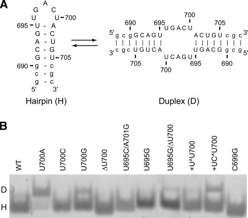
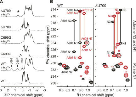
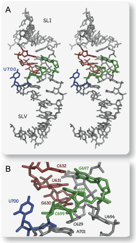
Substrate recognition by the VS ribozyme involves a magnesium-dependent loop/loop interaction between the SLI substrate and the SLV hairpin from the catalytic domain. Recent NMR studies of SLV demonstrated that magnesium ions stabilize a U-turn loop structure and trigger a conformational change for the extruded loop residue U700, suggesting a role for U700 in SLI recognition. Here, we kinetically characterized VS ribozyme mutants to evaluate the contribution of U700 and other SLV loop residues to SLI recognition. To help interpret the kinetic data, we structurally characterized the SLV mutants by NMR spectroscopy and generated a three-dimensional model of the SLI/SLV complex by homology modeling with MC-Sym. We demonstrated that the mutation of U700 by A, C, or G does not significantly affect ribozyme activity, whereas deletion of U700 dramatically impairs this activity. The U700 backbone is likely important for SLI recognition, but does not appear to be required for either the structural integrity of the SLV loop or for direct interactions with SLI. Thus, deletion of U700 may affect other aspects of SLI recognition, such as magnesium ion binding and SLV loop dynamics. As part of our NMR studies, we developed a convenient assay based on detection of unusual (31)P and (15)N N7 chemical shifts to probe the formation of U-turn structures in RNAs. Our model of the SLI/SLV complex, which is compatible with biochemical data, leads us to propose novel interactions at the loop I/loop V interface.


Figures

FIGURE 1.
FIGURE 1.
Substrate recognition by the Neurospora VS ribozyme. (A) Primary and secondary structures of the catalytic domain of the VS ribozyme. For this study, the AvaI ribozyme was used; it includes a 5′-TAIL with the sequence: 5′-gggaaagcuGCGG-3′ (Guo and Collins 1995). (B) The catalytic domain recognizes the SLI substrate (subdivided in Ia and Ib) via a loop/loop interaction involving residues 630–632 of SLI and residues 697–699 of SLV. Upon interaction with SLV, the SLI RNA with the wild-type sequence undergoes a structural change from an unshifted to a shifted conformation; only the later allows cleavage of the SLI internal loop at the site indicated by an arrowhead. (C) Preshifted mutant substrates, such as SLI(T), are efficiently cleaved by the VS catalytic domain (Andersen and Collins 2000; Zamel and Collins 2002). Wild-type and mutant nucleotides are represented by upper- and lowercase, respectively. Specific SLI and SLV nucleotides are color coded for easy identification in subsequent figures.
FIGURE 2.
FIGURE 2.
NMR structures of the SLV loop determined in the absence (left) and presence (right) of magnesium ions (Campbell and Legault 2005; Campbell et al. 2006), showing the large conformational change of U700 (blue). The dotted lines represent two hydrogen bonds that are characteristic of canonical U-turn structures, the A698 N7 to U696 2′OH (blue) and the U696 H3 to A698 3′-phosphate interactions (red). For simplicity, only heavy atoms are shown.
FIGURE 3.
FIGURE 3.
SLV loop mutants characterized kinetically and structurally in this study. Sequence and secondary structure of (A) SLV with U700 mutations and deletion, (B) SLV with mutations of the loop-closing base pair, (C) SLV with insertions of nucleotides between residues C699 and U700, and (D) SLV with a loop mutation that is expected to disrupt the loop I/loop V interaction (Rastogi et al. 1996) and also serves as an experimental control.
FIGURE 4.
FIGURE 4.
Kinetic analysis of the U700G Rz mutant. (A) Cleavage of 32P-labeled SLI(T) by the U700G Rz is performed at 30°C in the presence of 25 mM MgCl2 under single turnover conditions (excess enzyme [37.5 nM] over substrate [0.25 nM]). Denaturing polyacrylamide gel electrophoresis shows the amount of substrate (S) and product (P) at various time points of the reaction. (B) The natural log of the fraction of remaining substrate is plotted against time (see Materials and Methods). The data are fit to a single exponential and the first order rate constant is extracted by linear regression (kobs = absolute value of the slope = 0.12 min−1 and R2 = 0.9999). (C) Cleavage reactions with the U700G Rz are performed at various ribozyme concentrations and the value of kobs is plotted against ribozyme concentration. At low ribozyme concentration, there is a linear relationship between kobs and ribozyme concentration. The data are fit to a single exponential and the kcat/KM is extracted by linear regression (kcat/KM = value of the slope = 3.3 min−1μM−1 and R2 = 0.9988).
FIGURE 5.
FIGURE 5.
Conformation of SLV RNA fragments. (A) Primary and secondary structures of the wild-type SLV RNA fragment used for NMR studies in its hairpin (H) and duplex (D) conformations. (B) Native gel electrophoresis of the wild-type (WT) and mutant SLV RNAs. The RNAs were loaded at a concentration of 20 μM in the presence of 20 mM MgCl2.
FIGURE 6.
FIGURE 6.
NMR studies of SLV loop mutants. (A) 1D 1H-decoupled 31P spectra of wild-type (WT), C699G, and ΔU700 SLV RNA fragments in the absence and presence of 20 mM MgCl2 (+Mg2+). For the wild-type SLV in the presence of Mg2+, the most downfield-shifted resonances correspond to the 3′-phosphate of the residues indicated (Campbell et al. 2006). (B) Superpositions of a selected region from 2D 1H-15N long-range HMQC spectra recorded in the absence (black peaks) and presence (red peaks) of 20 mM MgCl2 for the wild-type (WT; left) and ΔU700 RNAs (right). The adenine H2-N1 and H2-N3 correlations of a given adenine can be easily identified based on the common H2 frequency and are linked by a line in the spectra. For the wild-type SLV, the A698 N7-H8 cross-peaks are indicated. In A and B, the asterisks point to a downfield-shifted 31P or upfield-shifted N7 resonance of an SLV mutant that appears in the presence of Mg2+.
FIGURE 7.
FIGURE 7.
(A) Stereo view of a structural model of the SLI/SLV complex. The selected model is a representative of the seven structures obtained from homology modeling with MC-Sym. For simplicity, only heavy atoms are shown. (B) Close up of the model shown in A.

Similar articles

Cited by

References

    1. Andersen, A., Collins, R.A. Rearrangement of a stable RNA secondary structure during VS ribozyme catalysis. Mol. Cell. 2000;5:469–478. - PubMed
    1. Andersen, A.A., Collins, R.A. Intramolecular secondary structure rearrangement by the kissing interaction of the Neurospora VS ribozyme. Proc. Natl. Acad. Sci. 2001;98:7730–7735. - PMC - PubMed
    1. Beattie, T.L., Collins, R.A. Identification of functional domains in the self-cleaving Neurospora VS ribozyme using damage selection. J. Mol. Biol. 1997;267:830–840. - PubMed
    1. Beattie, T.L., Olive, J.E., Collins, R.A. A secondary-structure model for the self-cleaving region of Neurospora VS RNA. Proc. Natl. Acad. Sci. 1995;92:4686–4690. - PMC - PubMed
    1. Cabello-Villegas, J., Tworowska, I., Nikonowicz, E.P. Metal ion stabilization of the U-turn of the A37 N6-dimethylallyl-modified anticodon stem–loop of Escherichia coli tRNAPhe . Biochemistry. 2004;43:55–66. - PubMed

Publication types