. 2007:372:505-28.
doi: 10.1007/978-1-59745-365-3_35.

Yeast mitochondrial transcriptomics

Affiliations

Yeast mitochondrial transcriptomics

Mathilde Garcia et al. Methods Mol Biol. 2007.

Abstract

Although 30 years ago it was strongly suggested that some cytoplasmic ribosomes are bound to the surface of yeast mitochondria, the mechanisms and the raison d'être of this process are not understood. For instance, it is not perfectly known which of the several hundred nuclearly encoded genes have to be translated to the mitochondrial vicinity to guide the import of the corresponding proteins. One can take advantage of several modern methods to address a number of aspects of the site-specific translation process of messenger ribonucleic acid (mRNA) coding for proteins imported into mitochondria. Three complementary approaches are presented to analyze the spatial distribution of mRNAs coding for proteins imported into mitochondria. Starting from biochemical purifications of mitochondria-bound polysomes, we describe a genomewide approach to classify all the cellular mRNAs according to their physical proximity with mitochondria; we also present real-time quantitative reverse transcription polymerase chain reaction monitoring of mRNA distribution to provide a quantified description of this localization. Finally, a fluorescence microscopy approach on a single living cell is described to visualize the in vivo localization of mRNAs involved in mitochondria biogenesis.


Figures
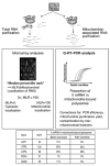
Fig. 1
Fig. 1
Biochemical methods to analyze the spatial distribution of mRNAs coding for proteins imported into mitochondria and example of results for ACT1 (contamination marker), ATP2, ATP3 (mitochondria-associated RNA), COX 4, and COX6 (nonmitochondria-associated RNA).
Fig. 2
Fig. 2
The distribution of mitochondrial location analyses can be skewed. (A) Example of distribution of the Cy5/Cy3 fluorescence ratios (Rf) that can be obtained from a standard global gene expression experiment. Both repressed and activated genes are expected; the distribution is similar to a normal distribution. (B) Example of distribution of Rf that can be obtained from microarray experiments comparing total RNAs and mitochondria-associated RNAs. Only RNAs enriched in the Cy5 channel are expected: the distribution is skewed by enriched RNA. Finally, single-cell fluorescent in situ hybridization (FISH) experiments (Subheadings 3.11. −3.15.) represent a necessary in vivo complement of the two preceding methods. Messenger ribonucleoproteins (mRNP) cytoplasmic localization can be directly observed by fluorescence microscopy, either in live cells using green fluorescent protein reporter proteins (9) or in fixed cells where endogenous mRNAs can be detected by FISH. New developments in probe design and in fluorophore chemistry allow routine detection of single molecules of mRNA within their cellular environment (10,11). FISH is particularly well suited or dissecting the different mechanisms governing mRNA localization in yeast (12). The protocol we present, adapted from refs. and , is designed to simultaneously compare the special distribution of different mRNAs relative to each other or to the mitochondria. FISH is particularly adapted in this case because mitochondria can be unambiguously detected using probes directed to the mitochondrial ribosomal RNAs (rRNAs), allowing for the simultaneous detection of mRNAs and mitochondrion in a single step. As an example, we show the simultaneous detection of the mitochondria-addressed ATP2 mRNA compared to the YRA1 mRNA, which is used as a nonlocalized control.
Fig. 3
Fig. 3
Mitochondria and localization of mRNA molecules. (A)–(D) Single-plane distribution of ATP2 mRNA: (A) Phase; (B) nucleus and mitochondria detected by DAPI staining; (C) ATP2 mRNA detected by FISH using Cy3-labeled probes. Arrows show the position of DAPI-stained cytoplasmic structures that can be difficult to discriminate from the very bright nucleus. (E)–(M) simultaneous detection of (E) DAPI, (F) the mitochondrial rRNA (Cy3.5), (G) ATP2 (Cy5), and (H) YRA1 (Cy3). Three consecutive planes were merged. The simultaneous detection of three different fluorophores requires the use of narrow-band filters, resulting in a noticeable diminution of the signal-to-noise ratios (compare G and C) (I) phase; (J) merge, DAPI in light gray (green) from E and mitochondrial rRNA from F; (K) merge mitochondrial rRNA in dark grey (red) from F and ATP2 mRNA in light gray (green) from G; (L) merge mitochondrial rRNA in dark gray (red) from F and YRA1 mRNA in light gray (green) from H; (M) 3D representation of the cellular volume (same cells as in E–L) ATP2 mRNA and mitochondrial rRNA are made green (Cy5) and purple (Cy3.5), respectively, and they appear as elongated intermingled gray structures. YRA1 mRNA can be observed in red (Cy3); it appears as round, single, gray structures. (N) Same as M showing the variability of ATP2addressing from cell to cell (compare M and N). Scale bar: 1 μm.

References

    1. Schatz G, Dobberstein B. Common principles of protein translocation across membranes. Science. 1996;271:1519–1526. - PubMed
    1. Neupert W. Protein import into mitochondria. Annu Rev Biochem. 1997;66:863–917. - PubMed
    1. Kellems RE, Allison VF, Butow RA. Cytoplasmic type 80S ribosomes associated with yeast mitochondria. IV. Attachment of ribosomes to the outer membrane of isolated mitochondria. J Cell Biol. 1975;65:1–14. - PMC - PubMed
    1. Verner K. Co-translational protein import into mitochondria: an alternative view. Trends Biochem Sci. 1993;18:366–371. - PubMed
    1. Suissa M, Schatz G. Import of proteins into mitochondria. Translatable mRNAs for imported mitochondrial proteins are present in free as well as mitochondria-bound cytoplasmic polysomes. J Biol Chem. 1982;257:13,048–13,055. - PubMed

Publication types

MeSH terms

LinkOut - more resources