. 2008 Mar;7(3):249-57.
doi: 10.1016/j.cmet.2008.01.005.

Insulin, cGMP, and TGF-beta signals regulate food intake and quiescence in C. elegans: a model for satiety

Affiliations

Insulin, cGMP, and TGF-beta signals regulate food intake and quiescence in C. elegans: a model for satiety

Young-jai You et al. Cell Metab. 2008 Mar.

Abstract

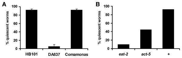
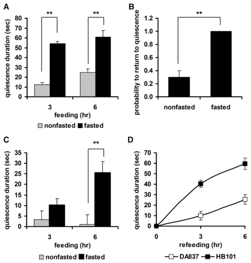
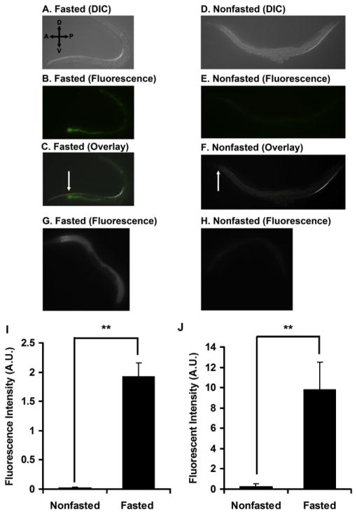
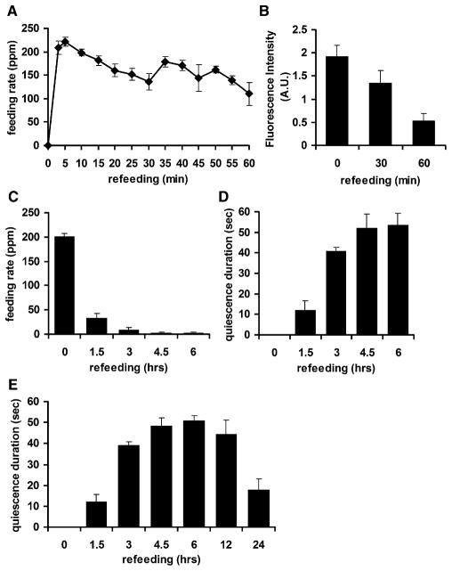
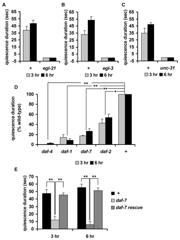
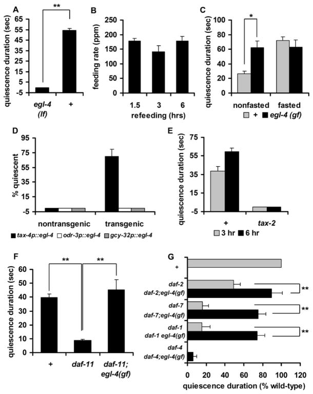
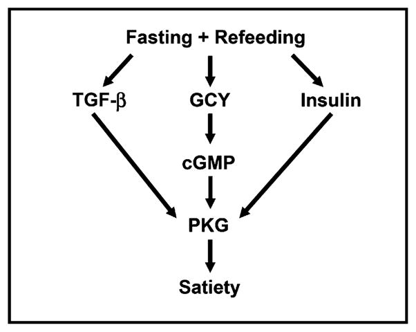
Despite the prevalence of obesity and its related diseases, the signaling pathways for appetite control and satiety are not clearly understood. Here we report C. elegans quiescence behavior, a cessation of food intake and movement that is possibly a result of satiety. C. elegans quiescence shares several characteristics of satiety in mammals. It is induced by high-quality food, it requires nutritional signals from the intestine, and it depends on prior feeding history: fasting enhances quiescence after refeeding. During refeeding after fasting, quiescence is evoked, causing gradual inhibition of food intake and movement, mimicking the behavioral sequence of satiety in mammals. Based on these similarities, we propose that quiescence results from satiety. This hypothesized satiety-induced quiescence is regulated by peptide signals such as insulin and TGF-beta. The EGL-4 cGMP-dependent protein kinase functions downstream of insulin and TGF-beta in sensory neurons including ASI to control quiescence in response to food intake.


Figures

Figure 1
Figure 1. Quiescence Is Induced by High-Quality Food and Is Dependent on Nutritional Status of Animals
(A) Quiescence (shown as percent quiescent worms) is dependent on food quality. Values are the mean ± SEM of two independent experiments. In each experiment, 20 wild-type worms were tested for each type of food. (B) Quiescence is dependent on nutrition, as shown by the effect of eat-2 and act-5 mutations. Twenty wild-type worms (+) and 20 of each type of mutant were tested on HB101.
Figure 2
Figure 2. Enhancement of Quiescence by Fasting and Refeeding
(A) Quiescence duration of fasted and refed worms is longer than that of non-fasted worms. Duration of quiescence was compared for fasted (black bars, n = 13) and nonfasted (gray bars, n = 19) groups after 3 or 6 hr of feeding with HB101. (B) Fasted and refed worms have a higher probability to return to quiescence than nonfasted worms. The probability to return to quiescence after nose touch (see Experimental Procedures) was compared between fasted (n = 10) and nonfasted groups (n = 11). (C) Fasting can induce quiescence on low-quality food. Duration of quiescence was compared for fasted (black bars, n = 20) and nonfasted (gray bars, n = 20) groups after 3 and 6 hr of feeding with DA837. (D) Induction of quiescence by low-quality food lags induction of quiescence by high-quality food. Duration of quiescence was compared for worms refed with HB101 (■) and worms refed with DA837 (□) after 3 or 6 hr of refeeding. Values are the mean ± SEM. **p < 0.001.
Figure 3
Figure 3. Fasting Enhances Food Intake during the Initial Period of Refeeding
(A) Differential interference contrast (DIC) image of a worm fasted for 12 hr and refed for 5 min on GFP-expressing HB101. (B) Fluorescence image of the worm in (A). (C) Overlay of (A) and (B). The arrow indicates strong GFP signal in the proximal intestine. (D) DIC image of a nonfasted worm fed for 5 min on GFP-expressing HB101. (E) Fluorescence image of the worm in (D). (F) Overlay of (D) and (E). The arrow indicates weak GFP signal in the proximal intestine. (G) Fluorescence image of a worm fasted for 12 hr and refed for 15 min on HB101 mixed with BODIPY dye. (H) Fluorescence image of a nonfasted worm fed for 15 min on HB101 mixed with BODIPY dye. (I) Quantification of images from ten fasted and ten nonfasted worms fed on GFP-expressing HB101, expressed in arbitrary units (A.U.) of fluorescence. Data are from one of three independent experiments. (J) Quantification of images from five fasted and five nonfasted worms fed for 15 min on HB101 mixed with BODIPY dye, expressed in arbitrary units (A.U.) of fluorescence. Data are from one of three independent experiments. Values are the mean ± SEM. **p < 0.001.
Figure 4
Figure 4. Quiescence Resembles the Behavioral Sequences of Satiety in Mammals
(A) Decrease in feeding rate (measured in pumps per minute [ppm]) over the time course of refeeding. Feeding rates of each of five wild-type worms were measured every 5 min for the first hour of refeeding. (B) Decrease in food intake as measured by GFP intensity. (C) Decrease in feeding rate (measured in ppm) over the time course of refeeding after 12 hr of fasting. Feeding rates of each of 20 wild-type worms were measured at each of the indicated time points (0, immediately after worms were transferred to food; 1.5, 1.5 hr after transfer, etc.) (D) Increase in quiescence duration over the time course of refeeding. The quiescence durations of ten worms were measured at the indicated time points after transfer to food. (E) Recovery of normal activity begins after 12 hr of refeeding. The x axis is not linear. Some points are replotted from (D). Values are the mean ± SEM.
Figure 5
Figure 5. Peptide Signals Including Insulin and TGF-β Mediate Quiescence after Fasting and Refeeding
In (A), (B), and (C), the x axis crosses the y axis at y = −5 so that the data for some mutants are visible. (A) Quiescence duration of egl-21 mutants. egl-21 mutants failed to show quiescence after 3 (gray bars) or 6 (black bars) hr of refeeding. (B) Quiescence duration of egl-3 mutants. egl-3 mutants failed to show quiescence after 3 (gray bars) or 6 (black bars) hr of refeeding. Four different alleles of egl-3 (nu349, gk238, n729, and nr2090) were tested, and all failed to show quiescence. The data from nr2090 are shown. (C) Quiescence duration of unc-31 mutants. unc-31 mutants failed to show quiescence after 3 (gray bars) or 6 (black bars) hr of refeeding. (D) Quiescence durations (expressed as a percent of wild-type) of daf-2 and TGF-β signaling mutants after 3 (gray bars) or 6 (black bars) hr of refeeding (see Experimental Procedures). (E) Expression of daf-7 in ASI neurons rescues the daf-7 quiescence defect. Values are the mean ± SEM. *p < 0.005, **p < 0.001. + indicates wild-type.
Figure 6
Figure 6. Insulin, cGMP, and TGF-β Signaling Regulate Quiescence through PKG in TAX-2/4-Expressing Neurons
In (A), (D), and (E), the x axis crosses the y axis at y = −5 so that the data for some mutants are visible. (A) Quiescence duration of egl-4 loss-of-function mutants (lf) after 3 hr of refeeding. Eleven egl-4(lf) mutants and thirteen wild-type (+) worms were used for the fasting/refeeding experiment. (B) Feeding rates (measured in ppm) of egl-4(lf) mutants at the indicated time points during refeeding (n = 11). (C) Quiescence duration of egl-4 gain-of-function mutants (gf) after 3 hr of isolation. For the nonfasted data, 19 nonfasted wild-type (+) and 20 nonfasted egl-4(gf) mutants were used. For the fasted data, 11 wild-type worms and 16 egl-4(gf) mutants were fasted and refed. (D) Expression of egl-4 under the control of a tax-4 promoter (black bars) rescued the egl-4 quiescence behavior defect after 6 hr of refeeding, whereas expressing egl-4 under the control of either an odr-3 (white bars) or a gcy-32 (gray bars) promoter did not. (E) Mutations in tax-2 abolish quiescence. tax-2 failed to show quiescence after 3 (gray bars) or 6 (black bars) hr of refeeding. Two different alleles of tax-2 (p671 and p694) were tested, and both failed to show quiescence. The data from p694 are shown. (F) daf-11 is an upstream guanylyl cyclase (GCY) regulating EGL-4 in quiescence. Quiescence durations of daf-11 and daf-11; egl-4(gf) double mutants. (G) Quiescence durations of TGF-β signaling mutants (gray bars) and double mutants with egl-4(gf) (black bars) calculated as percent of wild-type (see Experimental Procedures for SEM estimation). Values are the mean ± SEM. *p < 0.005, **p < 0.001.
Figure 7
Figure 7. Insulin, cGMP, and TGF-β Signaling Regulate Quiescence through PKG
Quiescence after fasting and refeeding is controlled by a signaling pathway in which PKG (EGL-4) is activated by insulin, cGMP, and TGF-β pathways.

References

    1. Anika SM, Houpt TR, Houpt KA. Insulin as a satiety hormone. Physiol Behav. 1980;25:21–23. - PubMed
    1. Ann K, Kowalchyk JA, Loyet KM, Martin TF. Novel Ca2+-binding protein (CAPS) related to UNC-31 required for Ca2+-activated exocytosis. J Biol Chem. 1997;272:19637–19640. - PubMed
    1. Antin J, Gibbs J, Holt J, Young RC, Smith GP. Cholecystokinin elicits the complete behavioral sequence of satiety in rats. J Comp Physiol Psychol. 1975;89:784–790. - PubMed
    1. Ashrafi K, Chang FY, Watts JL, Fraser AG, Kamath RS, Ahringer J, Ruvkun G. Genome-wide RNAi analysis of Caenorhabditis elegans fat regulatory genes. Nature. 2003;421:268–272. - PubMed
    1. Avery L. The genetics of feeding in Caenorhabditis elegans. Genetics. 1993;133:897–917. - PMC - PubMed

Publication types

MeSH terms