Comparative Study
. 2008 Aug 31;138(2):380-391.
doi: 10.1016/j.pain.2008.01.017. Epub 2008 Mar 7.

Sensitization of primary afferents to mechanical and heat stimuli after incision in a novel in vitro mouse glabrous skin-nerve preparation

Affiliations
Comparative Study

Sensitization of primary afferents to mechanical and heat stimuli after incision in a novel in vitro mouse glabrous skin-nerve preparation

Ratan K Banik et al. Pain. .

Abstract

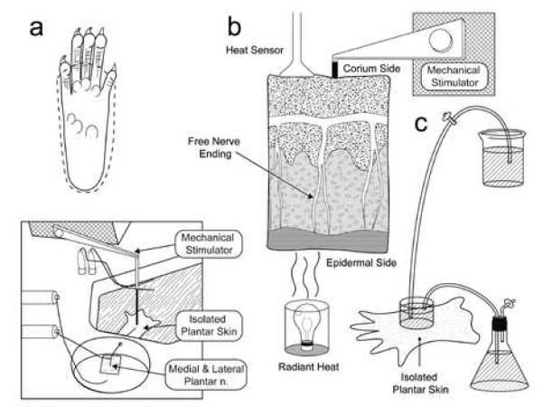
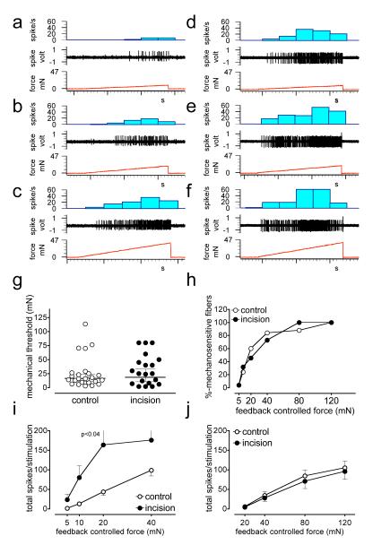
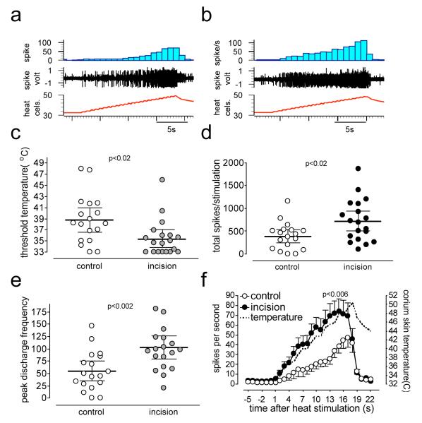
In this study, we recorded activity from afferent fibers innervating the mouse plantar skin, the same region evaluated in pain behavior experiments. We compared responses of afferents from incised and unincised hind paw skin. The plantar skin together with attached medial and lateral plantar nerves was dissected until they could be completely removed intact and placed in an organ bath chamber continuously perfused with oxygenated Kreb's solution with the temperature maintained at 32 degrees C. Afferent nerve activities to feedback-controlled mechanical and heat stimuli and cooling were recorded. Eighty-five single Adelta- and C-fiber afferents were recorded, 42 from control and the remainder from incised animals. A greater proportion of C-fibers (11/34) from incised skin had spontaneous activity than in the unincised preparation (2/32). The mechanical thresholds of both Adelta- and C-fiber units were not different between control and incised groups but the responses to suprathreshold mechanical stimulation were increased in low threshold Adelta- and C-fibers. The greatest change in heat sensitivity was apparent when multi-fiber total activity was measured; threshold was reduced, total spikes were greater and the peak discharge frequency was increased. In summary, feedback-controlled stimulation identified mechanical sensitization after incision in an in vitro preparation. Few fibers were excited by cooling. Heat sensitization of primary afferents was more prominent when activities of unclassified afferents are included. The preparation allows us to study afferent function of the same tissue that is examined for in vivo pain behavior assays in mice.


Figures

Figure 1
Figure 1. Experimental set up
A. above, dissection of rat plantar skin is done along the dotted line with medial and lateral plantar nerves. Below is a schematic of organ bath chamber and nerve-skin preparation. B. Computer controlled heat stimulation is given on the epidermal side skin, temperature was recorded by sensor in the corium side (dermis). Computer controlled mechanical stimulation is given in the corium side of skin. C. rapid exchange of ice cold Kreb’s solution onto receptive field produced cooling to 12 to 16°C.
Figure 2
Figure 2. A-δ fiber responses to mechanical stimuli
a-c, digitized oscilloscope tracings of an A-δ fiber responsive to mechanical stimuli. Single action potentials were recorded from fine filaments teased from medial or lateral plantar nerves of control mice while the receptive field was stimulated by a feedback controlled force stimulator. Three consecutive recordings show increasing responses to the ascending series of ramp shaped force stimuli starting from zero to 10 (a), 20 (b), and 40 (c) mN. The stimulus duration of each pulse was 5 sec and they were delivered at 60s intervals. d-f, digitized oscilloscope tracing of an A-δ fiber responsive to mechanical stimuli recorded from mice incised one day before recording (d, 10 mN, e, 20mN and f, 40mN). g-h, comparison of mechanical response thresholds between control and incised mice. The afferents are separated into two groups based on their mechanical response thresholds. Low threshold afferents produced responses by less than 10mN stimuli (i) while the threshold of high threshold afferents were above 10mN. Total number of spikes generated during ascending series of force pulses is compared in low (i) and high threshold (>10 mN) (j) afferents recorded from incised and control mice. P<0.04 vs control, Bonferroni’s post-hoc test.
Figure 3
Figure 3. C-fiber responses to mechanical stimuli
a-c, digitized oscilloscope tracings of a C-fiber responsive to mechanical stimuli. Three consecutive recording show increasing responses to the ascending series of ramp shaped force stimuli (a, 10 mN, b, 20mN and C, 40mN). d-f, digitized oscilloscope tracing of a C-fiber responsive to mechanical stimuli recorded from mice incised in the plantar hind paw one day before recording (d, 10 mN, e, 20mN and f, 40mN). g-h, comparison of mechanical response thresholds between control and incised mice. Comparison of total evoked spikes in the low (i) and high (j) threshold afferents to suprathreshold mechanical stimuli. . P<0.01 vs control, Bonferroni’s post-hoc test.
Figure 4
Figure 4. C-fiber responses to heat stimuli
Digitized oscilloscope tracings of C-fibers responsive to heat stimuli recorded from control (a) and incised (b) mice. c shows the threshold temperature for activation of C-fibers in single fiber analyses and d is the total spikes counted during heat stimulation. e shows peak discharge frequency during heat responses. P<0.03, unpaired t-test.
Figure 5
Figure 5. Heat responses after incision in multi-fiber analyses
Digitized oscilloscope tracings of multi-fiber responses to heat stimuli recorded from control (a) and incised (b) mice. In some recordings, in addition to mechanically identified single C-fiber, previously unidentified, heat responsive units were present. c shows the threshold temperature for activation of afferents in the multi-unit analyses and d is the total spikes counted during heat stimulation. e is the peak discharge frequency of afferents during heat response. The stimulus-response curve was drawn by averaging responses in one-second bins and by counting the total activity of fiber/(s) in that bin (f).
Figure 6
Figure 6. Cooling induced excitation
Digitized oscilloscope tracings of single fiber responses to cooling from incised mice. A is a recording from an A-δ fiber and b is the recording from a C-fiber. Two different response patterns are noted.

References

    1. Andrew D, Greenspan JD. Mechanical and heat sensitization of cutaneous nociceptors after peripheral inflammation in the rat. J Neurophysiol. 1999 Nov;82(5):2649–56. - PubMed
    1. Banik RK, Brennan TJ. Spontaneous discharge and increased heat sensitivity of rat C-fiber nociceptors are present in vitro after plantar incision. Pain. 2004 Nov;112(1-2):204–13. - PubMed
    1. Banik RK, Kozaki Y, Sato J, Gera L, Mizumura K. B2 receptor-mediated enhanced bradykinin sensitivity of rat cutaneous C-fiber nociceptors during persistent inflammation. Journal of neurophysiology. 2001 Dec;86(6):2727–35. - PubMed
    1. Banik RK, Woo YC, Park SS, Brennan TJ. Strain and sex influence on pain sensitivity after plantar incision in the mouse. Anesthesiology. 2006 Dec;105(6):1246–53. - PubMed
    1. Brennan TJ, Zahn PK, Pogatzki-Zahn EM. Mechanisms of incisional pain. Anesthesiology clinics of North America. 2005 Mar;23(1):1–20. - PubMed

Publication types