. 2008 May;76(5):1979-91.
doi: 10.1128/IAI.01338-07. Epub 2008 Mar 3.

A novel sensor kinase-response regulator hybrid controls biofilm formation and type VI secretion system activity in Burkholderia cenocepacia

Affiliations

A novel sensor kinase-response regulator hybrid controls biofilm formation and type VI secretion system activity in Burkholderia cenocepacia

Daniel F Aubert et al. Infect Immun. 2008 May.

Abstract

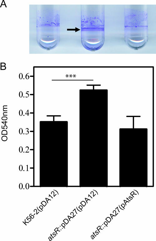
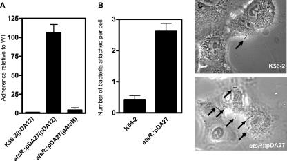
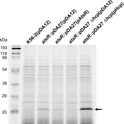
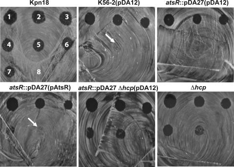
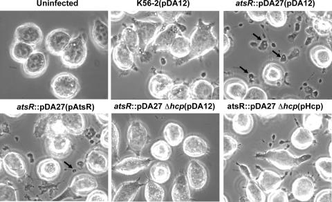
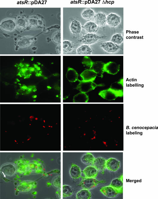
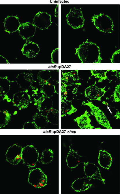
Burkholderia cenocepacia is an important opportunistic pathogen causing serious chronic infections in patients with cystic fibrosis (CF). Adaptation of B. cenocepacia to the CF airways may play an important role in the persistence of the infection. We have identified a sensor kinase-response regulator (BCAM0379) named AtsR in B. cenocepacia K56-2 that shares 19% amino acid identity with RetS from Pseudomonas aeruginosa. atsR inactivation led to increased biofilm production and a hyperadherent phenotype in both abiotic surfaces and lung epithelial cells. Also, the atsR mutant overexpressed and hypersecreted an Hcp-like protein known to be specifically secreted by the type VI secretion system (T6SS) in other gram-negative bacteria. Amoeba plaque assays demonstrated that the atsR mutant was more resistant to Dictyostelium predation than the wild-type strain and that this phenomenon was T6SS dependent. Macrophage infection assays also demonstrated that the atsR mutant induces the formation of actin-mediated protrusions from macrophages that require a functional Hcp-like protein, suggesting that the T6SS is involved in actin rearrangements. Three B. cenocepacia transposon mutants that were found in a previous study to be impaired for survival in chronic lung infection model were mapped to the T6SS gene cluster, indicating that the T6SS is required for infection in vivo. Together, our data show that AtsR is involved in the regulation of genes required for virulence in B. cenocepacia K56-2, including genes encoding a T6SS.


Figures

FIG. 1.
FIG. 1.
Biofilm assays performed with B. cenocepacia K56-2 (wild type [WT]) and DFA21 (the atsR::pDA27 mutant) containing the control plasmid pDA12 or pAtsR, as indicated in parentheses. Biofilms were quantified by crystal violet staining after 24 h of static incubation at 37°C. (A) The ring corresponding to robust biofilm formation characteristic of the atsR mutant is indicated by an arrow. (B) The extent of crystal violet retention by adherent bacteria was measured by OD540. Data shown are the means of three independent experiments done in triplicate. Error bars represent the standard deviations. Significant differences were determined using unpaired t tests. The three asterisks indicate that the P value is 0.0001.
FIG. 2.
FIG. 2.
Genetic organization and analysis of the gene cluster encoding AtsR in B. cenocepacia J2315 and K56-2. The direction of transcription of each gene is denoted by gray arrows. BCAM gene designations are according to a preliminary annotation of the B. cenocepacia J2315 genome (http://www.sanger.ac.uk/Projects/B_cenocepacia/). (A) Three-gene cluster located on chromosome 2 comprising BCAM0379 (atsR), BCAM0380, and BCAM0381. Black arrows represent genes located at the boundaries of the three-gene cluster containing atsR. Solid black bars indicate the regions analyzed by RT-PCR shown in panel B. (B) RT-PCR analysis of the intergenic regions of BCAM0381-BCAM 0380 and BCAM0380-atsR. Lanes: 1 to 3, RT, no RT, and DNA for BCAM0381-BCAM0380; 4 to 6, RT, no RT, and DNA for BCAM0380-atsR. White arrows indicate bands of expected PCR product size.
FIG. 3.
FIG. 3.
Adhesion assays. (A) Adhesion assay to a polystyrene surface of bacterial strains with the control vector pDA12 and the pAtsR plasmids, as indicated in parentheses. atsR indicates the DFA21 mutant. The adhesion values are shown relative to the value for the control strain, B. cenocepacia K56-2(pDA12), which was set as 1. (B) Adhesion assay on A549 human lung epithelial cells. A549 cells were incubated with B. cenocepacia K56-2 and B. cenocepacia DFA21 (indicated by atsR) for 1 h at 37°C, washed, and stained, and the adherent bacteria were enumerated. Data shown are the means of three independent experiments. Error bars represent the standard deviations. (C) Representative images of A549 cell adherence by B. cenocepacia K56-2 and B. cenocepacia DFA21 (atsR) are shown. Arrows point to adherent bacteria. WT, wild type.
FIG. 4.
FIG. 4.
Secretion assay. SDS-PAGE analysis of concentrated culture supernatants recovered from strains containing the control plasmid pDA12 or the plasmids pAtsR and pHcp, as indicated in parentheses. atsR indicates DFA21; atsR Δhcp indicates DFA28. Molecular mass markers in kDa are also indicated. The arrow points to the secreted Hcp polypeptide analysis of concentrated culture supernatants recovered from strains containing the control plasmid pDA12 or the plasmids pAtsR and pHcp, as indicated in parentheses. Molecular mass markers in kDa are also indicated. The arrow points to the position of secreted Hcp polypeptide. WT, wild type.
FIG. 5.
FIG. 5.
Genetic map of the T6SS gene cluster. The location of the transposon in the 34C6, 1A5, and 34C4 mutants, which are attenuated for survival in the rat agar bead model (23), is indicated by white circles. This cluster has been designated the bcs cluster for B. cenocepacia survival. The location and direction of transcription of genes are represented by arrows. Locus tags assigned by the Sanger Center are shown above and the bcs annotation of the genes is shown below. The genes icmF (BCAL0351; bcsB), clpV (BCAL0347; bcsF), and hcp (BCAL0343; bcsJ), which are characteristic of T6SSs in other bacteria, are shown.
FIG. 6.
FIG. 6.
Quantitative plaque assay. D. discoideum cells were applied as droplets onto a lawn of bacterial strains without plasmid or with the control vector pDA12 and pAtsR plasmids, as indicated in parentheses. atsR indicates DFA21; atsR Δhcp indicates DFA28; Δhcp indicates DFA27. The numbers of Dictyostelium cells applied were 50,000 (spot 1, as labeled for K. pneumoniae 18 [upper left]), 25,000 (spot 2), 12,500 (spot 3), 62,500 (spot 4), 3,125 (spot 5), 1,562 (spot 6), and 781 (spot 7); SorC buffer alone is shown for spot 8. After 3 days at 22°C, the ability of Dictyostelium cells to create plaques in the bacterial lawn was recorded. WT, wild type.
FIG. 7.
FIG. 7.
Phase-contrast microscopy of infected ANA-1 macrophages. The infections were performed at an MOI of 50:1 for 4 h with parental and mutant strains containing pDA12, pAtsR, or pHcp as indicated in parentheses. atsR indicates DFA21; atsR Δhcp indicates DFA28. Black arrows indicate vacuole-containing protrusions. WT, wild type.
FIG. 8.
FIG. 8.
Actin distribution of infected ANA-1 macrophages. Representative fluorescence micrographs of ANA-1 macrophages infected with strains DFA21 (shown as atsR) and DFA28 (shown as atsR Δhcp) at an MOI of 50:1 for 4 h are shown. Green fluorescence denotes filamentous actin. Red fluorescence denotes individual B. cenocepacia bacteria. The white arrow indicates a protrusion with a bacterium-containing vacuole. WT, wild type.
FIG. 9.
FIG. 9.
Actin distribution of infected ANA-1 macrophages. Representative confocal images of uninfected ANA-1 macrophages or ANA-1 macrophages infected with DFA21 (indicated as atsR) or DFA28 (indicated as atsR Δhcp) at an MOI of 50:1 for 4 h. Green fluorescence denotes filamentous actin. Red fluorescence denotes individual B. cenocepacia bacteria. The white arrow indicates a protrusion with a bacterium-containing vacuole.

References

    1. Bateman, A., L. Coin, R. Durbin, R. D. Finn, V. Hollich, S. Griffiths-Jones, A. Khanna, M. Marshall, S. Moxon, E. L. Sonnhammer, D. J. Studholme, C. Yeats, and S. R. Eddy. 2004. The Pfam protein families database. Nucleic Acids Res. 32D138-D141. - PMC - PubMed
    1. Chang, C., and R. C. Stewart. 1998. The two-component system. Regulation of diverse signaling pathways in prokaryotes and eukaryotes. Plant Physiol. 117723-731. - PMC - PubMed
    1. Coenye, T., and P. Vandamme. 2003. Diversity and significance of Burkholderia species occupying diverse ecological niches. Environ. Microbiol. 5719-729. - PubMed
    1. Cohen, S. N., A. C. Chang, and L. Hsu. 1972. Nonchromosomal antibiotic resistance in bacteria: genetic transformation of Escherichia coli by R-factor DNA. Proc. Natl. Acad. Sci. USA 692110-2114. - PMC - PubMed
    1. Corbett, C. R., M. N. Burtnick, C. Kooi, D. E. Woods, and P. A. Sokol. 2003. An extracellular zinc metalloprotease gene of Burkholderia cepacia. Microbiology 1492263-2271. - PubMed

Publication types

MeSH terms