. 2008 May;76(5):2002-7.
doi: 10.1128/IAI.01588-07. Epub 2008 Mar 3.

Mycolactone is responsible for the painlessness of Mycobacterium ulcerans infection (buruli ulcer) in a murine study

Affiliations

Mycolactone is responsible for the painlessness of Mycobacterium ulcerans infection (buruli ulcer) in a murine study

Junichiro En et al. Infect Immun. 2008 May.

Abstract

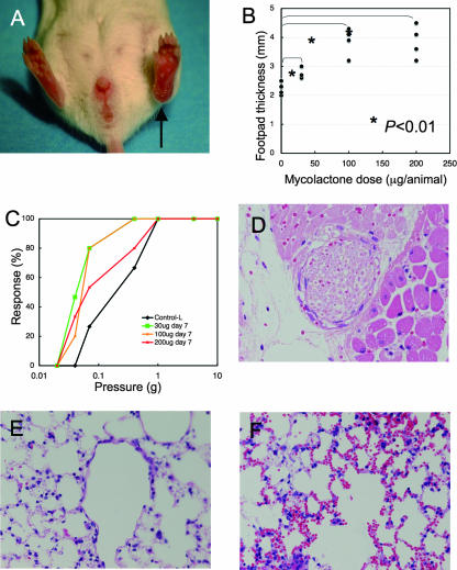
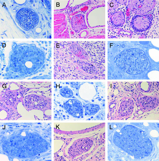
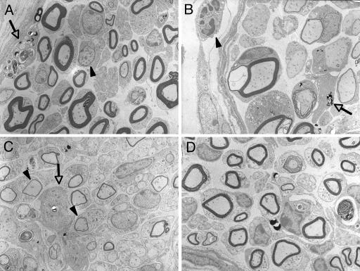
Buruli ulcer is a chronic skin disease caused by Mycobacterium ulcerans, which produces a toxic lipid mycolactone. Despite the extensive necrosis and tissue damage, the lesions are painless. This absence of pain prevents patients from seeking early treatment and, as a result, many patients experience severe sequelae, including limb amputation. We have reported that mice inoculated with M. ulcerans show loss of pain sensation and nerve degeneration. However, the molecules responsible for the nerve damage have not been identified. In order to clarify whether mycolactone alone can induce nerve damage, mycolactone A/B was injected to footpads of BALB/c mice. A total of 100 microg of mycolactone induced footpad swelling, redness, and erosion. The von Frey sensory test showed hyperesthesia on day 7, recovery on day 21, and hypoesthesia on day 28. Histologically, the footpads showed epidermal erosion, moderate stromal edema, and moderate neutrophilic infiltration up to day 14, which gradually resolved. Nerve bundles showed intraneural hemorrhage, neutrophilic infiltration, and loss of Schwann cell nuclei on days 7 and 14. Ultrastructurally, vacuolar change of myelin started on day 14 and gradually subsided by day 42, but the density of myelinated fibers remained low. This study demonstrated that initial hyperesthesia is followed by sensory recovery and final hypoesthesia. Our present study suggests that mycolactone directly damages nerves and is responsible for the absence of pain characteristic of Buruli ulcer. Furthermore, mice injected with 200 microg of mycolactone showed pulmonary hemorrhage. This is the first study to demonstrate the systemic effects of mycolactone.


Figures

FIG. 1.
FIG. 1.
Mycolactone injection into mouse footpad evoked swelling, hyperesthesia, nerve damage, and systemic pathology. Various amounts (30, 100, and 200 μg) of mycolactone A/B were injected into the left footpads of BALB/c mice. (A) Footpad swelling on day 7 after the injection of 100 μg (arrow). (B) Footpad thickness on day 7. Dose-dependent swelling was significant. (C) Sensory test showing hyperesthesia on day 7 (P = 0.02). (D) Nerve bundles showed a loss of Schwann cell nuclei and intraneural hemorrhage (H&E; magnification, ×168). (E) Lung without mycolactone injection (H&E, magnification, ×168). (F) Intra-alveolar hemorrhage in the lungs of mice injected with 200 μg of mycolactone into the footpad (H&E, magnification, ×168).
FIG. 2.
FIG. 2.
Sequential analysis of footpad thickness and sensation after mycolactone injection (100 μg). (A) Footpad thickness after mycolactone injection showed the peak swelling on day 14, with subsequent resolution. Sensory test showed significant hyperesthesia on day 7 (B, P = 0.017) and hyperesthetic tendency on day 14 (C, P = 0.081), normal sense on day 21 (D, P = 0.63), followed by significant hypoesthesia on day 28 (E, P = 0.0499) and hypoesthetic tendency on day 42 (F, P = 0.13).
FIG. 3.
FIG. 3.
Sequential analysis of footpad histopathology after mycolactone injection (100 μg). (A) Nerve of control mouse (only ethanol and 7H9 broth injected) showed no significant change. (B to D) On day 7, nerves of mycolactone-injected mice showed intraneural hemorrhage (B) and massive neutrophilic infiltration to the perineurium (C). (D) Epon section showing moderate loss of nerve fibers and myelin. (E and F) Nerves on day 14 showed intraneural inflammatory cell infiltration and hemorrhage. (G) Nerves on day 21. Mild infiltration inflammatory cell infiltration and vacuolar change were observed. (H) Vacuolar change of Schwann cells induced by mycolactone injection on day 21 was similar to M. ulcerans inoculation. (I) In the nerves on day 28, histological damage remained. (J) Thin myelins indicating remyelination were observed on day 28. (K) In the nerves on day 42, when sensory disturbance lasted, histological damage remained. (L) Mild fibrosis was observed on day 42. (A, D, F, H, J, and L [Epon, 1-μm sections, toluidine blue staining], magnification, ×340; B, C, E, G, I, and K [paraffin sections, H&E staining], magnification, ×157).
FIG. 4.
FIG. 4.
Ultrastructure of nerves after mycolactone injection. (A) Intraneural infiltration of lymphocytes (arrowhead) and macrophage (arrow) containing myelin debris on day 14 after mycolactone injection. (B) Vacuolar change of myelin (*) and macrophage (arrow) on day 21. (C) Thin myelins (arrows) indicate remyelination on day 28. D. Mild fibrosis observed on day 42. (Epon-embedded ultrathin sections stained by uranium and lead; magnification, ×1,960).

References

    1. Addo, P., E. Owusu, B. Adu-Addai, M. Quartey, M. Abbas, A. Doddo, and D. Ofori-Adjei. 2005. Findings from a Buruli ulcer mouse model study. Ghana Med. J. 3986-93. - PMC - PubMed
    1. Adusumilli, S., A. Mve-Obiang, T. Sparer, W. Meyers, J. Hayman, and P. L. Small. 2005. Mycobacterium ulcerans toxic macrolide, mycolactone modulates the host immune response and cellular location of M. ulcerans in vitro and in vivo. Cell Microbiol. 71295-1304. - PubMed
    1. Asiedu, K., W. Meyers, and P. Agbenorku. 2000. Clinical features and treatment, p. 37-38. In K. Asiedu, R. Scherpbier, and M. Raviglione (ed.), Buruli ulcer Mycobacterium ulcerans infection. World Health Organization, Geneva, Switzerland.
    1. Coutanceau, E., P. Legras, L. Marsollier, G. Reysset, S. T. Cole, and C. Demangel. 2006. Immunogenicity of Mycobacterium ulcerans Hsp65 and protective efficacy of a Mycobacterium leprae Hsp65-based DNA vaccine against Buruli ulcer. Microbes Infect. 82075-2081. - PubMed
    1. Dobos, K. M., P. L. Small, M. Deslauriers, F. D. Quinn, and C. H. King. 2001. Mycobacterium ulcerans cytotoxicity in an adipose cell model. Infect. Immun. 697182-7186. - PMC - PubMed

Publication types

LinkOut - more resources