. 2008 May;76(5):1992-2001.
doi: 10.1128/IAI.01221-07. Epub 2008 Mar 3.

Pseudomonas aeruginosa induces membrane blebs in epithelial cells, which are utilized as a niche for intracellular replication and motility

Affiliations

Pseudomonas aeruginosa induces membrane blebs in epithelial cells, which are utilized as a niche for intracellular replication and motility

Annette A Angus et al. Infect Immun. 2008 May.

Abstract

Pseudomonas aeruginosa is known to invade epithelial cells during infection and in vitro. However, little is known of bacterial or epithelial factors modulating P. aeruginosa intracellular survival or replication after invasion, except that it requires a complete lipopolysaccharide core. In this study, real-time video microscopy revealed that invasive P. aeruginosa isolates induced the formation of membrane blebs in multiple epithelial cell types and that these were then exploited for intracellular replication and rapid real-time motility. Further studies revealed that the type three secretion system (T3SS) of P. aeruginosa was required for blebbing. Mutants lacking either the entire T3SS or specific T3SS components were instead localized to intracellular perinuclear vacuoles. Most T3SS mutants that trafficked to perinuclear vacuoles gradually lost intracellular viability, and vacuoles containing those bacteria were labeled by the late endosomal marker lysosome-associated marker protein 3 (LAMP-3). Interestingly, mutants deficient only in the T3SS translocon structure survived and replicated within the vacuoles that did not label with LAMP-3. Taken together, these data suggest two novel roles of the P. aeruginosa T3SS in enabling bacterial intracellular survival: translocon-dependent formation of membrane blebs, which form a host cell niche for bacterial growth and motility, and effector-dependent bacterial survival and replication within intracellular perinuclear vacuoles.


Figures

FIG. 1.
FIG. 1.
A. Intracellular survival and replication of wild-type P. aeruginosa strains 6294, PAO1, and PAO1pscC::pCR2.1 (T3SS needle mutant) within human corneal epithelial cells after a 3-h incubation with 5 × 10⁵ CFU of bacteria prior to 1-h or 5-h gentamicin treatments (4-h and 8-h time points, respectively). Only wild-type strains survived and replicated. B. A similar experiment comparing strains PA103ΔexoUexoT::Tc and PA103pscJ::Tn5 (inoculum of 5 × 10⁶ CFU for PA103 mutants) or PAO1ΔexoSexoTexoY and PAO1ΔpopB (inoculum of 5 × 10⁵). *, P < 0.05 compared to 4-h time point for each strain; #, P < 0.05 compared to PAO1 parent strain (A) or PA103ΔexoUexoT::Tc isogenic mutant strain (B).
FIG. 2.
FIG. 2.
Intracellular location of P. aeruginosa wild-type PAO1 and T3SS mutants within human corneal epithelial cells after a 3-h incubation with 2 × 10⁷ CFU of bacteria followed by 1-h or 5-h gentamicin treatment (4-h and 8-h time points, respectively). PAO1 occupied spacious membrane blebs at 4 h (A) and 8 h (B). Uninfected cells maintained a healthy state at each time point (C and D, 4 and 8 h, respectively). A T3SS needle mutant, PAO1pscC::pCR2.1, was found in perinuclear vacuoles at 4 h (E) and 8 h (F). The mutant PAO1ΔexoSexoTexoY was not found in blebs or vacuoles at 4 h (G) but was found in vacuoles at 8 h (H). The translocon mutant, PAO1ΔpopB, also localized to vacuoles at each time point (I and J, 4 and 8 h, respectively). See also Video S1 in the supplemental material for real-time microscopy of wild-type strain PAO1 swimming inside blebs in this cell line.
FIG. 3.
FIG. 3.
Intracellular location of T3SS mutants of P. aeruginosa strain PA103 after a 3-h incubation with human corneal epithelial cells (2 × 10⁷ CFU of bacteria) followed by 1-h or 5-h gentamicin treatment (4-h and 8-h time points, respectively). The double effector mutant PA103ΔexoUexoT::Tc was not seen in membrane blebs or vacuoles at 4 h (A) but occupied membrane blebs at 8 h (B). The needle mutant PA103pscJ::Tn5 was also not observed within perinuclear vacuoles at 4 h (C) but was located within these vacuoles at 8 h (D). See also Video S2 in the supplemental material for real-time microscopy of strain PA103ΔexoUexoT::Tc inside blebs in this cell line.
FIG. 4.
FIG. 4.
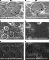
P. aeruginosa-induced membrane blebbing in other epithelial cell types. (A and B) Primary cultured rabbit corneal epithelial cells infected with 2 × 10⁶ CFU of clinical ocular isolate strain 6294 for 3 h prior to gentamicin treatment. Images were taken between 4 and 8 h postinfection. (C and D) Simian virus 40-immortalized rabbit corneal epithelial cells infected with 2 × 10⁷ CFU of strain PAK for 3 h prior to gentamicin treatment were then observed at the 4-h (C) and 8-h (D) time points. Human airway (alveolar) epithelial cells (A549) infected with 2 × 10⁷ CFU of strain PAO1 for 3 h prior to gentamicin treatment were then observed at 4 h (E) and 8 h (F). See also Videos S3 (6294), S4 (PAK), and S5 (PAO1) in the supplemental material for real-time microscopy of bacteria swimming inside blebs corresponding to the still images.
FIG. 5.
FIG. 5.
Intracellular colocalization of P. aeruginosa with the late endosome/lysosomal marker LAMP-3. Uninfected human corneal epithelial cells labeled with LAMP-3 (red) and DAPI (blue) (A). Wild-type PAO1 bacteria expressing GFP (green) were largely unassociated with LAMP-3 (B). The T3SS-null mutant PAO1ΔexsA expressing GFP was associated with cells in fewer numbers and colocalized with LAMP-3 adjacent to the nucleus (C). The T3SS translocon mutant PAO1ΔpopB was seen unassociated with LAMP-3 (D). All images were captured at 8 h postinfection with laser scanning confocal microscopy (see Materials and Methods). In each instance, cells were infected with 2 × 10⁷ CFU bacteria for 3 h before gentamicin treatment.
FIG. 5.
FIG. 5.
Intracellular colocalization of P. aeruginosa with the late endosome/lysosomal marker LAMP-3. Uninfected human corneal epithelial cells labeled with LAMP-3 (red) and DAPI (blue) (A). Wild-type PAO1 bacteria expressing GFP (green) were largely unassociated with LAMP-3 (B). The T3SS-null mutant PAO1ΔexsA expressing GFP was associated with cells in fewer numbers and colocalized with LAMP-3 adjacent to the nucleus (C). The T3SS translocon mutant PAO1ΔpopB was seen unassociated with LAMP-3 (D). All images were captured at 8 h postinfection with laser scanning confocal microscopy (see Materials and Methods). In each instance, cells were infected with 2 × 10⁷ CFU bacteria for 3 h before gentamicin treatment.

References

    1. Aktories, K., and J. T. Barbieri. 2005. Bacterial cytotoxins: targeting eukaryotic switches. Nat. Rev. 3397-410. - PubMed
    1. Alonso, A., and F. Garcia-del Portillo. 2004. Hijacking of eukaryotic functions by intracellular bacterial pathogens. Int. Microbiol. 7181-191. - PubMed
    1. Amano, A., I. Nakagawa, and T. Yoshimori. 2006. Autophagy in innate immunity against intracellular bacteria. J. Biochem. (Tokyo) 140161-166. - PubMed
    1. Baca, O. G., Y. P. Li, and H. Kumar. 1994. Survival of the Q fever agent Coxiella burnetii in the phagolysosome. Trends Microbiol. 2476-480. - PubMed
    1. Barbieri, J. T., and J. Sun. 2004. Pseudomonas aeruginosa ExoS and ExoT. Rev. Physiol. Biochem. Pharmacol. 15279-92. - PubMed

Publication types

LinkOut - more resources