. 2008 May;28(9):2980-95.
doi: 10.1128/MCB.02238-07. Epub 2008 Mar 3.

LRP-1 silencing prevents malignant cell invasion despite increased pericellular proteolytic activities

Affiliations

LRP-1 silencing prevents malignant cell invasion despite increased pericellular proteolytic activities

Stéphane Dedieu et al. Mol Cell Biol. 2008 May.

Abstract

The scavenger receptor low-density lipoprotein receptor-related protein 1 (LRP-1) mediates the clearance of a variety of biological molecules from the pericellular environment, including proteinases which degrade the extracellular matrix in cancer progression. However, its accurate functions remain poorly explored and highly controversial. Here we show that LRP-1 silencing by RNA interference results in a drastic inhibition of cell invasion despite a strong stimulation of pericellular matrix metalloproteinase 2 and urokinase-type plasminogen activator proteolytic activities. Cell migration in both two and three dimensions is decreased by LRP-1 silencing. LRP-1-silenced carcinoma cells, which are characterized by major cytoskeleton rearrangements, display atypical overspread morphology with a lack of membrane extensions. LRP-1 silencing accelerates cell attachment, inhibits cell-substrate deadhesion, and induces the accumulation, at the cell periphery, of abundant talin-containing focal adhesion complexes deprived of FAK and paxillin. We conclude that in addition to its role in ligand binding and endocytosis, LRP-1 regulates cytoskeletal organization and adhesive complex turnover in malignant cells by modulating the focal complex composition, thereby promoting invasion.


Figures

FIG. 1.
FIG. 1.
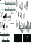
The short hairpin LRP-1 approach leads to specific LRP-1 silencing and to abrogation of LRP-1-mediated endocytosis in carcinoma cells. Total RNAs were purified from FTC133 wild-type (WT) cells, the control clonal cell line (shCTRL), two clonal cell lines that stably overexpress specific shRNAs for LRP-1 (shLRP-1-c1 and shLRP-1-c2), and siRNA-transfected cells (siCTRL and siLRP1). (A) The transcriptional level of LRP-1 was assessed by RT-PCR. GAPDH primers were used as a normalization control. (B) Whole-cell extracts from each cell line were subjected to immunoblot analysis with anti-LRP-1 antibody. β-Actin antibody was used for normalization. (C) Total RNAs were isolated from each cell line, and the expression of the interferon-stimulated proteins STAT1 and OAS was measured by RT-PCR. GAPDH primers were used as a control. (D) Each cell line was incubated for 30 min in serum-free medium containing FITC-labeled human α2M in the presence of 100 μM chloroquine. The specificity of internalization in WT and control cells was controlled by using a 100-fold higher concentration of the nonlabeled protein in a competition experiment (Compet.). The intracellular fluorescence was determined as described in Materials and Methods and expressed in relative units (R.U.) compared with the signal from WT cells. Each value is the mean ± standard deviation (SD) for three separate experiments. The gels and immunoblots are representative of three separate experiments. Numbers under the gels and immunoblots indicate the levels of induction (n-fold) by comparison with the WT (left panels) or siCTRL (right panels) cells. NS, differences from the WT are not significant; *, P < 0.01.
FIG. 2.
FIG. 2.
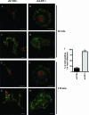
LRP-1 silencing mediates the increase of MMP-2- and uPA-dependent pericellular proteolysis and the stimulation of plasmin generation in human malignant cells. (A to G) WT, shCTRL, shLRP-1-c1, shLRP-1-c2, siCTRL, and siLRP-1 cells were cultured for 24 h in gelatin-coated dishes in the absence of serum. Gelatinolytic (A to C) and uPA-dependent (D to F) activities were evaluated by gelatin and gelatin-plasminogen zymography, respectively, with conditioned medium (C.M.) and membrane extracts (M.E.). Purified human MMP-2 (A) or uPA (D) was used as a positive control. Representative zymograms from three independent experiments are shown. Quantifications of the zymograms are presented in panels B, C, E, and F. (G) Plasmin generation was quantified by a colorimetric assay with the same samples. For panels B, E, and G, values were normalized by comparison to those obtained with WT cells. For panels C and F, values were normalized by comparison to those obtained with siCTRL cells. Results are presented in relative units, as means ± SD for triplicate wells from three separate experiments. NS, not significant; *, significantly different from the corresponding control (P < 0.01). (H) Control and LRP-1-silenced cells were subjected to in situ zymography, using Bodipy FL casein as a fluorogenic substrate, to detect pericellular proteolytic activities (green). Bars, 20 μm.
FIG. 3.
FIG. 3.
LRP-1 silencing contributes to maintaining high levels of the soluble proteinases MMP-2 and uPA secreted into the extracellular environment. (A) Serum-free conditioned medium from WT, shCTRL, shLRP-1-c1, shLRP-1-c2, siCTRL, and siLRP-1 cells was collected after 24 h. The proteins were subjected to Western blotting analysis using anti-uPA, anti-MMP-2, and anti-β-actin antibodies. Purified human MMP-2 and uPA were used as positive controls. (B) Total RNAs were isolated from these cells, and the expression of the MMP-2 and uPA genes was assessed by RT-PCR. GAPDH amplification was used as a control. Representative gels and immunoblots are presented. Numbers under the gels and immunoblots indicate the levels of induction (n-fold) by comparison with WT (left panels) or siCTRL (right panels) cells.
FIG. 4.
FIG. 4.
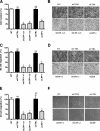
Carcinoma cell invasion and migration processes are altered to the same extent by LRP-1 silencing. Cell invasion and cell migration assays were carried out with WT, shCTRL, shLRP-1-c1, shLRP-1-c2, siCTRL, and siLRP-1 cells. (A and B) Tumor cell invasion was measured on Matrigel-coated Transwell membranes. (C and D) Three-dimensional cell migration was assessed by using uncoated filters. (E and F) Two-dimensional cell migration was determined by a wound-healing assay. Representative images are shown. Results for invasion assays were obtained from eight separate experiments, with each performed in triplicate, and results for migration assays were obtained from three separate experiments, with each performed in triplicate. Invasion and migration were determined by counting cells in eight random microscopic fields per well. Results are expressed as means ± SD after normalization by comparison with WT cells. NS, differences from WT were not significant; *, P < 0.01.
FIG. 5.
FIG. 5.
LRP-1 silencing stimulates the rate of carcinoma cell attachment and inhibits cell-substrate deadhesion. (A) shCTRL and shLRP-1-c1 cells were seeded onto gelatin-coated plates, and the nonadherent cells were discarded after 30, 60, 90, 120, or 180 min. For each cell type, results are expressed as percentages of adherent cells. (B) The same experiment was performed using siCTRL and siLRP-1 cells. (C) Control (shCTRL and siCTRL) and LRP-1-silenced (shLRP-1 and siLRP-1) cells were grown in gelatin-coated dishes for 24 h and subjected to trypsinization assay by incubating cells with 0.025% (wt/vol) trypsin for 10 min. For each cell type, results are expressed as percentages of detached cells. Each value is the mean ± SD for four separate experiments, with each performed in triplicate. NS, differences from corresponding control were not significant; *, P < 0.01.
FIG. 6.
FIG. 6.
LRP-1 silencing promotes human carcinoma cell spreading. shCTRL (A, C, E, and G) and shLRP-1-c1 (B, D, F, and H) cells were seeded onto gelatin-coated plates for 30 (A and B), 60 (C and D), 90 (E and F), or 120 (G and H) min and visualized by phase-contrast microscopy. Images are representative of three separate sets of cultures. Similar results were obtained with shLRP-1-c2 cells. Bars, 40 μm.
FIG. 7.
FIG. 7.
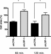
LRP-1-silenced carcinoma cells exhibit a twofold increased cell area compared to control cells. shCTRL and shLRP-1-c1 cells were seeded onto gelatin-coated plates for 60 or 120 min. Cell area was calculated with Image J software by using 200 isolated cells for each cell line in two independent experiments. The shCTRL cell area was scaled up to 100%. *, P < 0.01. Similar results were obtained with shLRP-1-c2 cells.
FIG. 8.
FIG. 8.
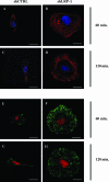
LRP-1-silenced carcinoma cells exhibit striking differences in focal adhesion organization. shCTRL (A, C, E, and G) and shLRP-1-c1 (B, D, F, and H) cells were plated onto gelatin-coated coverslips for 60 or 120 min. Cells were stained for actin filaments (red) (A to D) or alpha-actinin (green) (E to H), and nuclei were counterstained in blue (A to D) or red (E to H). Images are representative of three separate experiments. Similar results were obtained with shLRP-1-c2 cells. Bars, 20 μm.
FIG. 9.
FIG. 9.
Disruption of talin-containing focal complexes is inhibited in LRP-1-silenced carcinoma cells. shCTRL (A, C, E, and G) and shLRP-1-c1 (B, D, F, and H) cells were plated onto gelatin-coated coverslips for 60 or 120 min. Isolated cells (A, B, E, and F) and grouped cells (C, D, G, and H) were stained for talin (green), and nuclei were counterstained (red). Images are representative of three separate experiments. Bars, 20 μm. (I) Cells were assayed for the percentage of cells positive for focal adhesions. Two hundred fifty cells for each clonal cell line were evaluated from three separate experiments. *, P < 0.01. Similar results were obtained with shLRP-1-c2 cells.
FIG. 10.
FIG. 10.
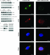
LRP-1 mediates the control of focal complex composition in malignant cells. (A and B) shCTRL and shLRP-1-c1 cells were plated onto gelatin-coated coverslips for 60, 120, or 180 min. Whole-cell extracts were subjected to Western blot analysis. (A) Anti-alpha-actinin and anti-talin antibodies were used. (B) Antipaxillin, antivinculin, anti-phospho-FAK (Y576/577), and anti-FAK antibodies were used. β-Actin labeling was used for normalization. (C) Immunoprecipitation of talin-containing focal complexes was performed (IP: anti-talin), and the immunocomplexes were immunoblotted (IB) by using antitalin, antipaxillin, and anti-FAK antibodies. Nonspecific IgG was used as a negative control for immunoprecipitation. Immunoblots are representative of three separate experiments. (D) Control and LRP-silenced cells were stained for paxillin (green) and p-FAK (red), and nuclei were counterstained (blue). Representative images are shown. Bars, 10 μm. Similar results were obtained with siLRP-1-transfected cells.

References

    1. Andreasen, P. A., R. Egelund, and H. H. Petersen. 2000. The plasminogen activation system in tumor growth, invasion, and metastasis. Cell Mol. Life Sci. 5725-40. - PMC - PubMed
    1. Barmina, O. Y., H. W. Walling, G. J. Fiacco, J. M. Freije, C. Lopez-Otin, J. J. Jeffrey, and N. C. Partridge. 1999. Collagenase-3 binds to a specific receptor and requires the low density lipoprotein receptor-related protein for internalization. J. Biol. Chem. 27430087-30093. - PubMed
    1. Boucher, P., M. Gotthardt, W. P. Li, R. G. Anderson, and J. Herz. 2003. LRP: role in vascular wall integrity and protection from atherosclerosis. Science 300329-332. - PubMed
    1. Bu, G., P. A. Morton, and A. L. Schwartz. 1992. Identification and partial characterization by chemical cross-linking of a binding protein for tissue-type plasminogen activator (t-PA) on rat hepatoma cells. A plasminogen activator inhibitor type 1-independent t-PA receptor. J. Biol. Chem. 26715595-15602. - PubMed
    1. Chazaud, B., S. Bonavaud, A. Plonquet, M. Pouchelet, R. K. Gherardi, and G. Barlovatz-Meimon. 2000. Involvement of the [uPAR:uPA:PAI-1:LRP] complex in human myogenic cell motility. Exp. Cell Res. 258237-244. - PubMed

Publication types

MeSH terms