. 2008 Mar 10;180(5):867-75.
doi: 10.1083/jcb.200706189. Epub 2008 Mar 3.

Cdk11 is a RanGTP-dependent microtubule stabilization factor that regulates spindle assembly rate

Affiliations

Cdk11 is a RanGTP-dependent microtubule stabilization factor that regulates spindle assembly rate

Hideki Yokoyama et al. J Cell Biol. .

Abstract

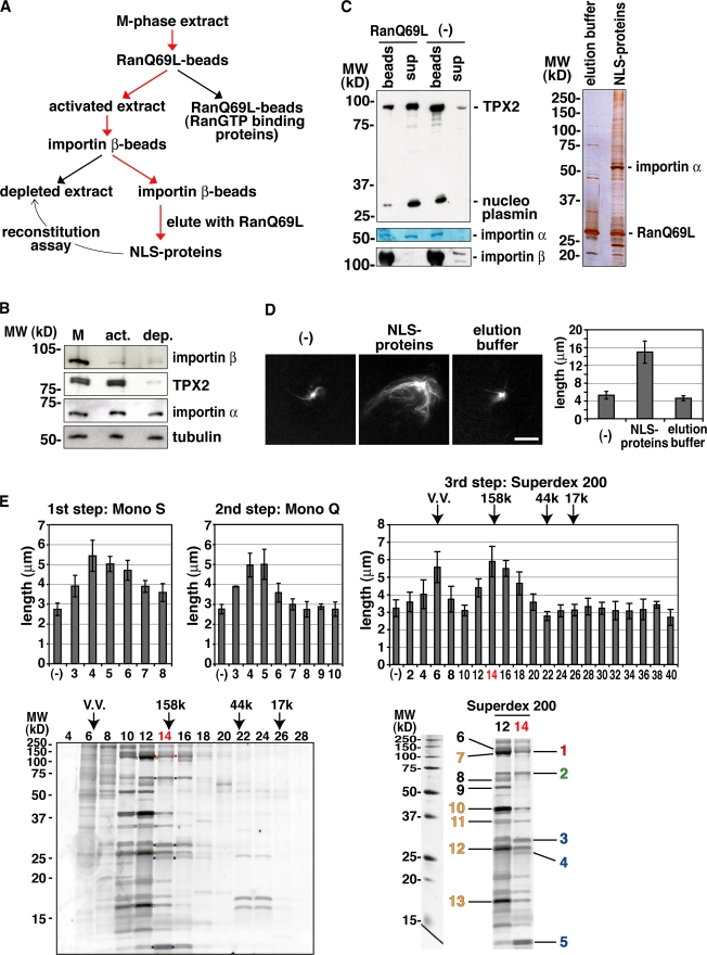
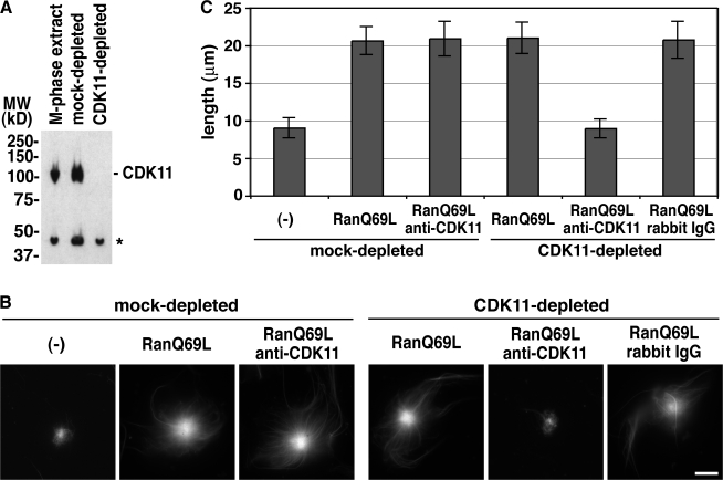
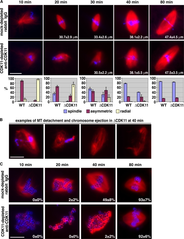
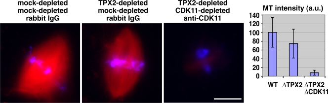
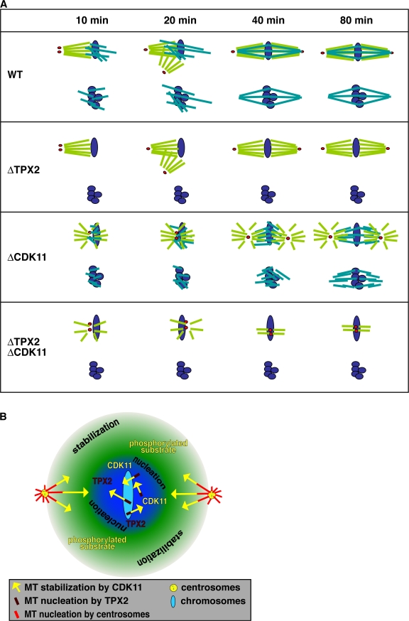
Production of Ran-guanosine triphosphate (GTP) around chromosomes induces local nucleation and plus end stabilization of microtubules (MTs). The nuclear protein TPX2 is required for RanGTP-dependent MT nucleation. To find the MT stabilizer, we affinity purify nuclear localization signal (NLS)-containing proteins from Xenopus laevis egg extracts. This NLS protein fraction contains the MT stabilization activity. After further purification, we used mass spectrometry to identify proteins in active fractions, including cyclin-dependent kinase 11 (Cdk11). Cdk11 localizes on spindle poles and MTs in Xenopus culture cells and egg extracts. Recombinant Cdk11 demonstrates RanGTP-dependent MT stabilization activity, whereas a kinase-dead mutant does not. Inactivation of Cdk11 in egg extracts blocks RanGTP-dependent MT stabilization and dramatically decreases the spindle assembly rate. Simultaneous depletion of TPX2 completely inhibits centrosome-dependent spindle assembly. Our results indicate that Cdk11 is responsible for RanGTP-dependent MT stabilization around chromosomes and that this local stabilization is essential for normal rates of spindle assembly and spindle function.


Figures

Figure 1.
Figure 1.
Purification of the RanGTP-dependent MT stabilization activity from Xenopus egg extract. (A) Purification strategy. (B) Immunoblot of the extract outlined in A. M, M-phase extract; act, activated extract; dep, depleted extract. The labeled proteins were detected by specific antibodies. (C) Elution of NLS proteins and endogenous importin α from the importin β column by RanQ69L and 500 mM NaCl. (left) After incubation, supernatant (sup) and beads were analyzed by immunoblotting (TPX2, nucleoplasmin, and importin β) or Coomassie staining (importin α). (right) Silver staining of proteins in elution buffer or the NLS protein fraction. Note that a major band (importin α) and various other bands (expected NLS proteins) were detected in addition to RanQ69L. (D) MT stabilization activity detected in the NLS protein fraction. The NLS protein fraction or elution buffer was incubated in the depleted extract containing centrosomes, anti-TPX2 antibodies, and Cy3-labeled tubulin (left). The MT length of the centrosomal asters was quantified (right), as shown in Fig. S1 A (available at http://www.jcb.org/cgi/content/full/jcb.200706189/DC1). (E) Purification of the MT stabilization activity from the NLS protein fraction. First step, Mono S; second step, Mono Q; third step, Superdex 200. Molecular mass standards (kD) are indicated. VV, void volume. The MT stabilization activity of purified fractions was assayed as described in Fig. 1 D. (bottom left) SDS-PAGE and Cypro ruby staining of the Superdex 200 fractions. Red boxes, Cdk11; green boxes, DKC1, NOP5, and XNOP56; blue boxes, small nuclear RNP proteins U2A′, U2B″, and Sm proteins; orange boxes, the processing of precursor complex. (bottom right) Proteins identified by mass spectrometry from the active stabilization fractions (Table I). The colored numbers correspond to the colored boxes in the bottom left panel. (D and E) Error bars represent SD. n > 20 asters; n > 3 experiments. Bar, 20 μm.
Figure 2.
Figure 2.
Cdk11 is responsible for RanGTP-dependent MT stabilization in M-phase extracts. (A) Depletion of Cdk11 from M-phase extracts. An M-phase extract was immunodepleted using control or anti–full-length Cdk11 antibodies, and the depletion efficiency was evaluated by immunoblotting. The asterisk indicates a cross-reacting band. (B) RanGTP-dependent MT stabilization assay. The mock- or Cdk11-depleted extracts were incubated with centrosomes, anti-TPX2 antibodies, and Cy3-labeled tubulin in the presence or absence of 12 μM RanQ69L, 0.22 mg/ml anti-Cdk11 antibodies, or rabbit IgG. (C) Quantification of the MT length assayed in B as described in Fig. S1 A (available at http://www.jcb.org/cgi/content/full/jcb.200706189/DC1). Error bars represent SD. n > 20 asters. This experiment was reproduced three times. Bar, 20 μm.
Figure 3.
Figure 3.
Cdk11-dependent MT stabilization is required for normal spindle assembly rate. (A and C) Representative structures observed in mock- or Cdk11-depleted extracts supplemented with 0.44 mg/ml rabbit IgG or anti-Cdk11 antibodies, respectively. Cycled spindles were assembled in the presence of Cy3-labeled tubulin (red), fixed at the indicated time points by squashing, and stained with Hoechst 33342 (blue). These experiments were reproduced three times. (A) Sperm spindle assembly. The numbers inside images represent the mean length of spindles. n > 30 spindles. The graphs below the images represent the percentage of structures observed according to the code indicated. n > 50 structures; n > 2 experiments. Error bars represent SD. WT, wild type. (B) Examples of abnormal sperm spindle structures observed in a Cdk11-inactivated extract at 40 min. They represent 36 ± 5% of all structures observed (47 ± 3% of spindles) and were not incorporated in the graph in A. (C) DNA bead spindle assembly. The numbers inside the images represent the percentages of bipolar spindles observed over the total number of structures counted. n > 50 structures; n > 2 experiments. Bars, 20 μm.
Figure 4.
Figure 4.
Cdk11 is essential for spindle assembly in the absence of TPX2. Representative spindle structures observed under the indicated conditions. Cycled sperm spindles were assembled in the presence of Cy3-tubulin (red), fixed at 80 min, centrifuged onto coverslips, and stained with Hoechst 33342 (blue). MT intensity in spindles was quantified using a macro. Error bars represent SD. n > 50 spindles. This experiment was reproduced three times. WT, wild type. Bar, 20 μm.
Figure 5.
Figure 5.
Schematic interpretation of the effects of Cdk11 on spindle MTs. (A) Spindle assembly around sperm nuclei and DNA beads. Sperm spindles: in wild-type (WT) extracts, both centrosomal and chromosomal MTs are stabilized by Cdk11 and contribute to spindle assembly. Initially, centrosomal MTs grow asymmetrically toward chromosomes through a Cdk11-dependent MT stabilization. In the absence of TPX2, centrosomal MTs still grow asymmetrically and form a bipolar spindle. In the absence of Cdk11, centrosomal MTs are not stabilized but interact with short chromosomal MTs. The MT populations become organized by cross-linking motors. When both TPX2 and Cdk11 are inactivated, there are no chromosomal MTs, and centrosomal MTs are too short and dynamic to be organized by cross-linking motors. Red dots, centrosomes; purple ovals, chromosomes; light green, centrosomal MTs; blue green, TPX2-dependent chromosomal MTs. DNA bead spindles: in wild-type extract, spindles form exclusively from TPX2 nucleated chromosomal MTs. In the absence of TPX2, no MTs are formed. In the absence of Cdk11, short and dynamic MTs assemble and spindle assembly is slowed down, but cross-linking motors finally organize them into a normal spindle containing short MTs. When both TPX2 and Cdk11 are inactivated, no MTs are nucleated. Purple, DNA beads; blue green, TPX2-dependent chromosomal MTs. (B) Hypothetical Cdk11 stabilization gradient. Cdk11 is released from importins and activated around chromosomes approximately over the same area as other NLS molecules like TPX2. However, its phosphorylated substrate that stabilizes MTs can diffuse further away before being dephosphorylated by the global opposing phosphatase. Yellow arrows indicate the increased length of MTs caused by the local activity of Cdk11.

References

    1. Bastiaens, P., M. Caudron, P. Niethammer, and E. Karsenti. 2006. Gradients in the self-organization of the mitotic spindle. Trends Cell Biol. 16:125–134. - PubMed
    1. Berke, J.D., V. Sgambato, P.P. Zhu, B. Lavoie, M. Vincent, M. Krause, and S.E. Hyman. 2001. Dopamine and glutamate induce distinct striatal splice forms of Ania-6, an RNA polymerase II-associated cyclin. Neuron. 32:277–287. - PubMed
    1. Burbank, K.S., T.J. Mitchison, and D.S. Fisher. 2007. Slide-and-cluster models for spindle assembly. Curr. Biol. 17:1373–1383. - PubMed
    1. Carazo-Salas, R.E., G. Guarguaglini, O.J. Gruss, A. Segref, E. Karsenti, and I.W. Mattaj. 1999. Generation of GTP-bound Ran by RCC1 is required for chromatin-induced mitotic spindle formation. Nature. 400:178–181. - PubMed
    1. Carazo-Salas, R.E., O.J. Gruss, I.W. Mattaj, and E. Karsenti. 2001. Ran-GTP coordinates regulation of microtubule nucleation and dynamics during mitotic-spindle assembly. Nat. Cell Biol. 3:228–234. - PubMed

Publication types

MeSH terms