. 2008 Mar 1;22(5):640-53.
doi: 10.1101/gad.1632608.

Methylation of RUNX1 by PRMT1 abrogates SIN3A binding and potentiates its transcriptional activity

Affiliations

Methylation of RUNX1 by PRMT1 abrogates SIN3A binding and potentiates its transcriptional activity

Xinyang Zhao et al. Genes Dev. .

Abstract

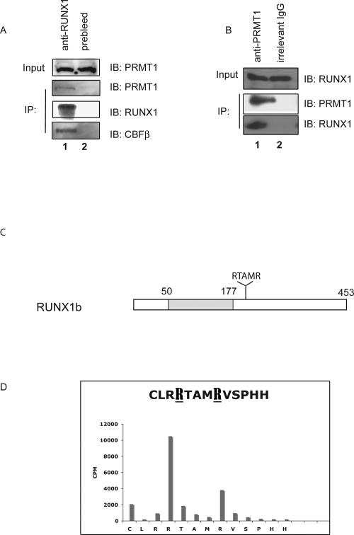
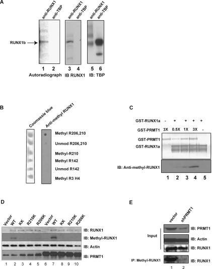
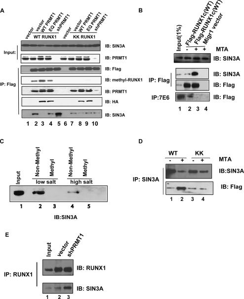
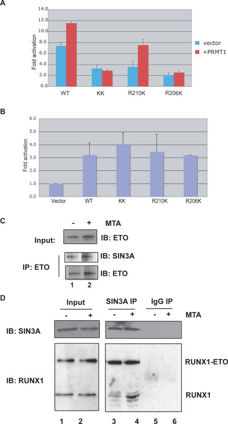
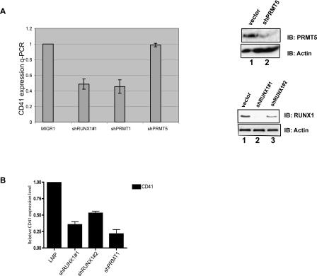
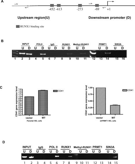
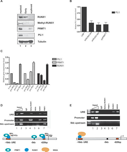
RUNX1/AML1 is required for the development of definitive hematopoiesis, and its activity is altered by mutations, deletions, and chromosome translocations in human acute leukemia. RUNX1 function can be regulated by post-translational modifications and protein-protein interactions. We show that RUNX1 is arginine-methylated in vivo by the arginine methyltransferase PRMT1, and that PRMT1 serves as a transcriptional coactivator for RUNX1 function. Using mass spectrometry, and a methyl-arginine-specific antibody, we identified two arginine residues (R206 and R210) within the region of RUNX1 that interact with the corepressor SIN3A and are methylated by PRMT1. PRMT1- dependent methylation of RUNX1 at these arginine residues abrogates its association with SIN3A, whereas shRNA against PRMT1 (or use of a methyltransferase inhibitor) enhances this association. We find arginine-methylated RUNX1 on the promoters of two bona fide RUNX1 target genes, CD41 and PU.1 and show that shRNA against PRMT1 or RUNX1 down-regulates their expression. These arginine methylation sites and the dynamic regulation of corepressor binding are lost in the leukemia-associated RUNX1-ETO fusion protein, which likely contributes to its dominant inhibitory activity.


Figures

Figure 1.
Figure 1.
The endogenous RUNX1 and PRMT1 proteins physically interact in HEL cells. (A) An anti-RUNX1 antibody was used to immunoprecipitate PRMT1 from HEL cell extracts, using preimmune rabbit serum as control. As a positive control, CBFβ was detected among the anti-RUNX1 immunoprecipitated proteins. (B) An anti-PRMT1 murine antibody was used to immunoprecipitate RUNX1 from HEL cell extracts, using an irrelevant murine IgG antibody as negative control. (C) Schematic diagram of RUNX1b, showing the position of the Runt domain shaded and the RTAMR methylation site; the numbers on top correspond to the amino acid position. (D) Results of Edman degradation of an in vitro methylated synthetic peptide that contains amino acids 203–215 of RUNX1. Two sites in RUNX1 that are arginine-methylated by recombinant PRMT1 are identified.
Figure 2.
Figure 2.
RUNX1 is arginine-methylated in vivo. (A) HEL cells were treated with protein synthesis inhibitors before [3H-methyl]-methionine was added to the medium for labeling. The cell lysate was then immunoprecipitated with an anti-RUNX1 antibody (lanes 1,3) or an irrelevant (anti-TBP) antiserum (lanes 2,4). Methylated RUNX1 is shown in the left panel, and the middle and right panels are immunoprecipitation controls for the two antibodies used. (B) The anti-methyl-arginine-specific RUNX1 antibody efficiently recognizes the R206 and R210 diasymmetrically methylated peptide, but not the unmethylated peptide, the R210-only asymmetrically methylated peptide, the R142 asymmetrically methylated peptide, or the R3 asymmetrically methylated H4 histone tail peptide in a dot blot analysis. (C) The anti-methyl-arginine RUNX1 antibody recognizes recombinant RUNX1 only after in vitro methylation by recombinant GST-PRMT1. The top panel is the PVDF membrane stained for protein, and the bottom panel is a Western blot assay performed with the anti-methyl-arginine-RUNX1 antibody. (D) The wild-type RUNX1 protein, but not the R206K and R210K mutant proteins, is doubly arginine-methylated in HeLa cells, as detected using the anti-methyl-arginine RUNX1 antibody. Lane 1 is HeLa cells transfected with the empty pCDNA3 vector, whereas lanes 25 contain overexpressed wild-type RUNX1c or the various R-to-K RUNX1 mutants. Lane 6 contains HeLa cells that overexpress PRMT1 alone, and lanes 710 contain overexpressed RUNX1c and overexpressed PRMT1. The overexpression of PRMT1 leads to greater methylation of RUNX1 wild-type protein (cf. lanes 2 and 7), without changing the level of RUNX1 expression. Similarly, overexpression of RUNX1c did not change the level of PRMT1 (cf. lanes 1 and 2). (E) Knocking down PRMT1 in HEL cells with shRNA reduces the amount of arginine-methylated RUNX1 but not the total amount of RUNX1 protein. Lane 1 is the vector-integrated HEL cells. Lane 2 shows the decrease in PRMT1 and methyl RUNX1 in HEL cells that stably express PRMT1 shRNA. Actin levels serve as the loading control.
Figure 3.
Figure 3.
PRMT1 regulates the association between RUNX1 and SIN3A by methylating RUNX1 at R206 and R210. (A) The association of SIN3A with wild-type RUNX1 but not the R206KR210K mutant is reciprocally affected by increasing or decreasing the level of PRMT1. Furthermore, PRMT1 enzymatic activity is required to decrease the association of RUNX1 with SIN3A. Anti-Flag antibody-coated agarose was used to immunoprecipitate Flag-tagged RUNX1 proteins from HeLa cells overexpressing wild-type or mutant forms of RUNX1 with wild-type PRMT1 (lanes 3,8), enzymatic dead PRMT1 (EQ) (lanes 4,9), or shRNA directed against PRMT1 (lanes 5,10). Equal loading in all lanes is shown by the SIN3A IB (in the top panel), the expression of RUNX1 protein is shown in the “IB: Flag” panel, the amount of RTAMR-methylated RUNX1 protein is shown in the “IB: methyl-RUNX1” panel, the level of PRMT1 in the anti-Flag immunoprecipitate is shown in the “IB: PRMT1” panel, the level of overexpressed PRMT1 in the anti-Flag immunoprecipitate is shown in the “IB: HA” panel, and the amount of SIN3A in the anti-Flag immunoprecipitate is shown in the bottom “IB: SIN3A” panel. (B) The methylation inhibitor MTA was added to HEL cells for 15 h, as indicated. More SIN3A is bound to Flag-RUNX1c (wild type) when MTA is present (cf. lanes 2 and 3 in A). (C) An arginine-methylated RUNX1 peptide does not pull down the SIN3A complex, whereas the unmethylated RUNX1 peptide does. (Lane 1, input) Ten percent nuclear extract, used for the peptide pulldown assay. Peptide-bound proteins were washed with 60 mM NaCl (lanes 2,3) or 100 mM NaCl (lanes 4,5). SIN3A protein is detected by an anti-SIN3A antibody. (D) In contrast, the association between SIN3A and the R206K R210K RUNX1 mutant protein (shown as KK in this figure) is not altered by MTA treatment. HEL cells expressing wild-type or mutant Flag-tagged RUNX1 were subjected to immunoprecipitation using an anti-SIN3A monoclonal antibody. The immunoprecipitated proteins were detected by anti-SIN3A and anti-Flag antibodies. (E) PRMT1 is primarily responsible for regulating the association of RUNX1 with SIN3A. Knocking down the level of PRMT1 by shRNA in a stably transfected HEL cell line increases the binding of RUNX1 with SIN3A. Input (lane 1) contains 10% of the amount of HEL cell extract used for the immunoprecipitation studies, whereas lanes 2 and 3 contain empty vector stably transduced and PRMT1-directed shRNA stably transduced cells, respectively. The immunoprecipitation was done using an anti-RUNX1 antibody; the coimmunoprecipitated protein was detected using the anti-SIN3A monoclonal antibody G11.
Figure 4.
Figure 4.
The in vivo association of RUNX1 with SIN3A (and presumably HDACs) regulates its transcriptional activity in HEL cells. (A) RUNX1 R-to-K mutants weakly activate the IL-3 promoter in HeLa cells compared with wild-type RUNX1, and the KK and R206K mutant forms do not respond to PRMT1 expression. The transfections were performed in the presence (in red) or absence (in blue) of PRMT1. (B) The addition of TSA restores transactivating properties to the RUNX1 R-to-K mutants on the IL-3 promoter. (C) The addition of MTA does not change the amount of SIN3A bound to ETO. Lanes 1 and 2 both contain HEL cell proteins immunoprecipitated with an anti-ETO antibody. (D) The addition of MTA for 12 h to Kasumi-1 cells enhances the binding of SIN3A to RUNX1, but has no effect on SIN3A binding to RUNX1–ETO. (Lanes 3,4) Following immunoprecipitation of SIN3A using an anti-SIN3A monoclonal antibody, the coimmunoprecipitated RUNX1 and RUNX1–ETO proteins were detected using an anti-RUNX1 antibody in a Western blot assay. (Lanes 5,6) Normal mouse IgG is used as the control.
Figure 5.
Figure 5.
Expression of CD41, a direct target gene of RUNX1, is regulated by RUNX1 and PRMT1 in primary cells. (A) Knocking down RUNX1 or PRMT1 levels (but not PRMT5 levels) using shRNA reduces CD41 expression in HEL cells, as shown by real-time PCR. The ability of the PRMT5- and RUNX1-directed shRNAs to lower the level of PRMT5 and RUNX1 protein is shown on the right. (B) CD41 expression is reduced in CD34+ cord blood cells grown in early cytokine mix by knocking down RUNX1 or PRMT1 using shRNA-expressing retroviral vectors.
Figure 6.
Figure 6.
Assessment of transcriptional regulators bound to the CD41 promoter. (A) A schematic diagram of the CD41 promoter is shown, indicating the upstream (U) and downstream (D) regions. (B) RUNX1 (lanes 8,9), PRMT1 (lanes 13,14), and methyl RUNX1 (lanes 10,11) are sitting on the actively transcribed CD41 promoter (∼70 bp upstream of the start site) in HEL cells, but not on an ∼5-kb upstream region of the gene (which we use as a control for the ChIP). An anti-Pol II antibody (lanes 4,5) served as a positive control and normal IgG (lanes 6,7) served as a negative control for the PCR reactions. Lanes 1 and 12 show the 50-bp DNA sizing ladder. Input lanes contain 0.01% of genomic input DNA. The PCR products were resolved using a 20% 1× TBE PAGE gel. (C) Comparison of the effects of overexpressing RUNX1 in HEL cells that stably express shPRMT1 (right panel) versus the parental HEL cells (left panel). RUNX1 slightly increases CD41 expression in HEL cells, but it reduces CD41 expression in cells with low PRMT1 levels. (D) ChIP assays show that in the shPRMT1-expressing HEL cells, overexpression of RUNX1 leads to the recruitment of SIN3A to the CD41 promoter (lane 15) but not PRMT1 or methyl-RUNX1 (lanes 11,13). (Lane 6) Pol II is present (and possibly stalled) on the CD41 promoter. Lane 9 is the DNA sizing ladder.
Figure 7.
Figure 7.
Regulation of RUNX1, PRMT1, and methyl RUNX1 levels during differentiation of human CD34+ cells in liquid culture. (A) Runx1 is arginine-methylated by PRMT1 during myeloid differentiation. CD34+ cells isolated from human cord blood cells were cultured under three different cytokine conditions: early (progenitor maintaining) mix, promyeloid differentiation mix, or proerythroid differentiation mix. PRMT1 protein is up-regulated under promyeloid and proerythroid differentiation conditions, RUNX1 and PU.1 protein are up-regulated with promyeloid differentiation mix and RUNX1 and PU.1 protein are both down-regulated in the proerythroid mix. (B) PU.1 expression is down-regulated in CD34+ cells grown in promyeloid mix following the reduction of RUNX1 or PRMT1 levels. (C) Real-time PCR results for the level of PRMT1, RUNX1, and PU.1 mRNA expression. (E) Early mix; (pro-Meg) megakaryocytic promoting mix; (pro-Ery) erythroid-promoting mix; (pro-Mye) myeloid-promoting mix. PU.1 expression is down-regulated in the pro-Ery and pro-Meg mix. RUNX1 is down-regulated in the pro-Ery mix. Both are up-regulated in the pro-Mye mix. (D) ChIP assays show that cells with higher PU.1 expression (grown in promyeloid mix) have both PRMT1 and methylated RUNX1 bound to the PU.1 promoter region and the URE. Only PRMT1 is bound to a region 5 kb upstream of the PU.1 transcriptional start site. (E) ChIP assays show that in the pro-Meg mix, SIN3A and RUNX1, but not PRMT1 or methyl RUNX1, are found on the URE. This accompanies the marked drop in PU.1 expression seen in these cells. Input lanes contain 0.01% and 0.05% of input genomic DNA used in panels D and E.

References

    1. An W., Kim J., Roeder R.G. Ordered cooperative functions of PRMT1, p300, and CARM1 in transcriptional activation by p53. Cell. 2004;117:735–748. - PubMed
    1. Bedford M.T., Richard S. Arginine methylation an emerging regulator of protein function. Mol. Cell. 2005;18:263–272. - PubMed
    1. Boisvert F.M., Cote J., Boulanger M.C., Richard S. A proteomic analysis of arginine-methylated protein complexes. Mol. Cell. Proteomics. 2003;2:1319–1330. - PubMed
    1. Bruhn L., Munnerlyn A., Grosschedl R. ALY, a context-dependent coactivator of LEF-1 and AML-1, is required for TCRα enhancer function. Genes & Dev. 1997;11:640–653. - PubMed
    1. Chakraborty S., Sinha K.K., Senyuk V., Nucifora G. SUV39H1 interacts with AML1 and abrogates AML1 transactivity. AML1 is methylated in vivo. Oncogene. 2003;22:5229–5237. - PubMed

Publication types

MeSH terms