. 2008 Mar 1;14(5):1470-7.
doi: 10.1158/1078-0432.CCR-07-1450.

HMGA1 is a molecular determinant of chemoresistance to gemcitabine in pancreatic adenocarcinoma

Affiliations

HMGA1 is a molecular determinant of chemoresistance to gemcitabine in pancreatic adenocarcinoma

Siong-Seng Liau et al. Clin Cancer Res. .

Erratum in

Abstract

Purpose: HMGA1 proteins are architectural transcription factors that are overexpressed by pancreatic adenocarcinomas. We previously have shown that RNA interference targeting the HMGA1 gene may represent a potential chemosensitizing strategy in pancreatic adenocarcinoma cells. In this study, we tested the hypothesis that HMGA1 promotes chemoresistance to gemcitabine in pancreatic cancer cells.

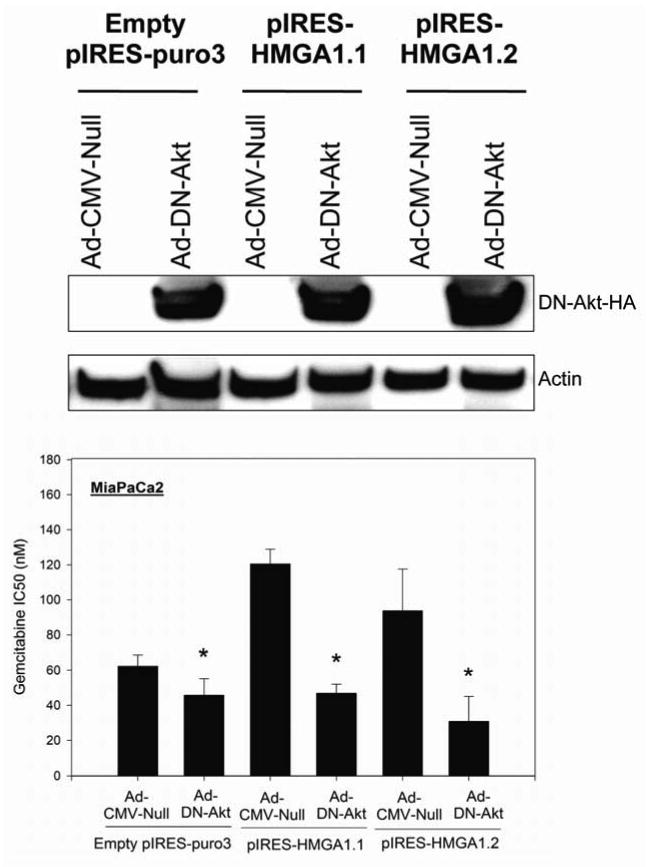
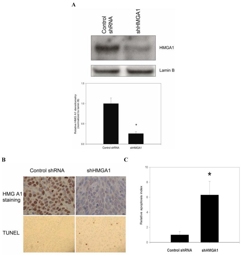
Experimental design and results: Stable short hairpin RNA-mediated HMGA1 silencing in BxPC3 and MiaPaCa2 cells promoted chemosensitivity to gemcitabine, with reductions in gemcitabine IC(50) and increases in gemcitabine-induced apoptosis and caspase-3 activation. In contrast, forced HMGA1 overexpression in MiaPaCa2 cells promoted chemoresistance to gemcitabine, with increases in gemcitabine IC(50) and reductions in gemcitabine-induced apoptosis and caspase-3 activation. Dominant negative Akt abrogated HMGA1 overexpression-induced increases in chemoresistance to gemcitabine. Finally, HMGA1 silencing promoted chemosensitivity to gemcitabine in vivo in a nude mouse xenograft model of pancreatic adenocarcinoma.

Conclusion: Our findings suggest that HMGA1 promotes chemoresistance to gemcitabine through an Akt-dependent mechanism. Targeted therapies directed at HMGA1 represent a potential strategy for ameliorating chemoresistance in pancreatic adenocarcinoma.


Figures

Fig. 1
Fig. 1. BxPC3: Lentivirus-mediated silencing of HMGA1 and chemosensitivity to gemcitabine
A, Following the generation of high titre lentivirus particles carrying shHMGA1, BxPC3 was transduced with lentivirus at MOI of 10 and stable transfectants were developed following selection with puromycin. Robust suppression of HMGA1 was achieved using lentivirus with a high degree of silencing of HMGA1. In BxPC3 cells, lentivirus-mediated shHMGA1 achieved almost complete silencing of HMGA1. Controls were stable transfectants developed using lentivirus carrying scrambled, non-targeting shRNA. B, The effects of lentivirus-mediated HMGA1 silencing on chemosensitivity to gemcitabine were assessed. When BxPC3 cells in which HMGA1 had been silenced were exposed to 1μM gemcitabine for 48 hours, they adopted a less healthy, speculated morphology as compared to control cells. Photomicrographs were taken using an inverted microscope at 40X magnification. C, Survival curves following exposure to 0-10μM gemcitabine were analyzed following MTS assay. Lentivirus-mediated stable HMGA1 silencing in BxPC3 cells shifted the survival curve to the left indicating an increased in chemosensitivity to gemcitabine, when compared to the controls. Correspondly, there was 4-fold reductions in the IC50 to gemcitabine with silencing of HMGA1 when compared to the controls. *P=0.001 versus control shRNA.
Fig 2
Fig 2. MiaPaCa2: Effects of lentivirus-mediated silencing and forced overexpression of HMGA1 on chemosensitivity to gemcitabine
MiaPaCa2 cells were transduced with shRNA lentivirus at MOI of 10 and stable transfectants were developed following selection with puromycin. Stable silencing of HMGA1 was confirmed by Western analysis (data not shown). Two previously characterized clones (pIRES-HMGA1.1 and 1.2) of MiaPaCa2 cells which stably overexpress HMGA1 were used in this study (19). Controls were cells stably transfected with empty pIRES-puro3 vector. A, The effects of modulating HMGA1 expression on chemosensitivity to gemcitabine in MiaPaCa2 cells was assessed. In MiaPaCa2 cells, lentivirus-mediated silencing of HMGA1 resulted in marked reductions in cellular viability as compared to controls when exposed to 1μM gemcitabine for 72 hours. In contrast, MiaPaCa2 cells with HMGA1 overexpression (pIRES-HMGA1.1 and 1.2) demonstrated increased viability following exposure to 1μM gemcitabine, when compared to empty pIRES-puro3 transfectants. Photomicrographs were taken using an inverted microscope at 40X magnification. B-C, Lentivirus-mediated RNA interference of HMGA1 in MiaPaCa2 cells resulted in increases in chemosensitivity to gemcitabine with shifting of survical curves to the left while overexpression of HMGA1 led increases in chemoresistance to gemcitabine with shifting of the survival curves to the right in both pIRES-HMGA1.1 and 1.2 clones, compared to their respective controls. D, Targeted suppression of HMGA1 using lentivirus-mediated shHMGA1 resulted in 2-fold reductions in IC50 compared to control (p=0.001 versus control), while overexpression of HMGA1 in pIRES-HMGA1.1 and 1.2 clones resulted in approximately 2.2 and 1.7-fold increases in IC50 to gemcitabine respectively (p=0.003 and p=0.006 versus empty pIRES-puro3 control). *P<0.05 versus control shRNA transfectants. **P<0.05 versus empty pIRES-puro3 transfectants.
Fig 3
Fig 3
A, Lentivirus-mediated silencing of HMGA1 promoted gemcitabine-induced apoptosis as assessed by flow cytometric analyses of YO-PRO-1/Propidium iodide-stained cells. Silencing of HMGA1 resulted in approximately 2-fold increases in the relative apoptotic rates in both BxPC3 and MiaPaCa2 cells following exposure to 1μM gemcitabine for 24 hours. *P=0.001 versus control shRNA. Representative flow cytometric images of three experiments are shown, with the apoptotic fractions being highlighted in triangles drawn. B, Forced overexpression of HMGA1 in pIRES-HMGA1.1 and 1.2 clones protected the cells from gemcitabine-induced apoptosis with approximately 70-80% reductions in relative apoptotic rates, as assessed by flow cytometry [P=0.001 (pIRES-HMGA1.1) and P=0.002 (pIRES-HMGA1.2) versus empty pIRES-puro3 control]. *P<0.05 versus empty pIRES-puro3 control. C, Relative caspase 3 activities were determined using a fluorometric caspase 3 substrate assay following exposure of cells to 1μM gemcitabine for 24 hours. Lentivirus-mediated silencing of HMGA1 promoted gemcitabine-induced caspase 3 activities. *P=0.008 (BxPC3) and P=0.005 (MiaPaCa2) versus control shRNA transfectants. D, As expected, overexpression of HMGA1 in pIRES-HMGA1.1 and 1.2 clones resulted in reductions in caspase 3 activation following exposure to 1μM gemcitabine for 24 hours, indicating protection from gemcitabine-induced caspase-mediated apoptosis. *P=0.001 versus empty pIRES-puro3 transfectants.
Fig 4
Fig 4
We have previously reported that in MiaPaCa2 cells, HMGA1 silencing reduces Akt phosphorylation whilst HMGA1 overexpression promotes Akt phosphorylation, and neither HMGA1 silencing nor overexpression have any impact on the level of expression of total Akt. To assess the role of Akt in mediating the HMGA1-induced chemoresistance, we transduced pIRES-HMGA1.1 and 1.2 clones with adenovirus carrying HA-tagged dominant negative Akt to examine its effects on chemoresistance. Transduction efficiency and expression of dominant negative Akt were assessed by Western blotting for hemagglutinin. Infection of pIRES-HMGA1.1 and 1.2 clones with adenovirus carrying dominant negative Akt (Ad-DN-Akt) resulted in significant reductions in IC50 to gemcitabine when compared to cells infected with control adenovirus (Ad-CMV-Null). Dominant negative Akt resulted in reductions in IC50 to gemcitabine in pIRES-HMGA1.1 and 1.2 clones to levels similar to parental MiaPaCa2 cells or empty pIRES-puro3 transfectants, indicating abrogation of the increased chemoresistance associated with HMGA1 overexpression. *P<0.05 versus control adenovirus (Ad-CMV-Null).
Fig 5
Fig 5
A, Stable silencing of HMGA1 promoted chemosensitivity to gemcitabine in vivo with evidence of tumor regression in nude mouse subcutaneous model. Mice (n=8 per group) were subcutaneously implanted with 2X10⁶ lentivirus-mediated stable transfectant BxPC3 cells (either shHMGA1 or control shRNA). Gemcitabine treatment was commenced in each group 14 days after implantation when the tumors were approximately 50mm3 in volume. Mice received gemcitabine (150 mg/kg) in 100 μL of PBS vehicle by twice-weekly i.p. injection. Subcutaneous tumor size was monitored weekly during the 6 weeks of treatment. Tumors with HMGA1 silencing showed evidence of regression in size during the treatment period while tumors in the control group continued to grow with time. Values are means (± SD). *P<0.05 versus control shRNA xenografts. B, Representative photograph of one mouse from each group is shown, with tumors located in their flanks. C, The explanted tumors at the end of the 6-week treatment period are shown. Of note, one of the mice in the shHMGA1 group had its tumor completely regress during the 6-week gemcitabine treatment period.
Fig 6
Fig 6
A, In vivo HMGA1 silencing was confirmed by Western blot analysis of nuclear extracts from explanted xenograft tumors. Densitometric values are means (± SD). *P=0.001 versus control shRNA xenografts. B-C, Immunohistochemistry of xenograft sections demonstrated little or absent staining for HMGA1 in the shHMGA1 xenografts, when compared to the control shRNA xenografts which showed intense staining for HMGA1. Photomicrograph of HMGA1 staining was obtained at 40X magnification. In the same sections, TUNEL staining was performed. In each tumor slide stained with TUNEL, the number of TUNEL-positive cells were counted in at least 5 randomly selected fields at 40X magnification. HMGA1 silencing led to significant increases in relative apoptotic index when compared to control shRNA xenografts. Representative tumor sections stained for TUNEL photographed at X20 magnification are shown. *P<0.001 versus control shRNA xenografts. Values are means (± SD).

Comment in

References

    1. Jemal A, Siegel R, Ward E. Cancer statistics, 2006. CA Cancer J Clin. 2006;56:106–30. - PubMed
    1. Sener SF, Fremgen A, Menck HR, Winchester DP. Pancreatic cancer: a report of treatment and survival trends for 100,313 patients diagnosed from 1985-1995, using the National Cancer Database. J Am Coll Surg. 1999;189:1–7. - PubMed
    1. Friedmann M, Holth LT, Zoghbi HY, Reeves R. Organization, inducible-expression and chromosome localization of the human HMG-I(Y) nonhistone protein gene. Nucleic Acids Res. 1993;21:4259–67. - PMC - PubMed
    1. Thanos D, Maniatis T. Virus induction of human IFN beta gene expression requires the assembly of an enhanceosome. Cell. 1995;83:1091–100. - PubMed
    1. Du W, Thanos D, Maniatis T. Mechanisms of transcriptional synergism between distinct virus-inducible enhancer elements. Cell. 1993;74:887–98. - PubMed

MeSH terms