. 2008 Mar 4;105(9):3262-7.
doi: 10.1073/pnas.0710897105. Epub 2008 Mar 3.

Cloning and identification of an oxytocin/vasopressin-like receptor and its ligand from insects

Affiliations

Cloning and identification of an oxytocin/vasopressin-like receptor and its ligand from insects

Elisabeth Stafflinger et al. Proc Natl Acad Sci U S A. .

Abstract

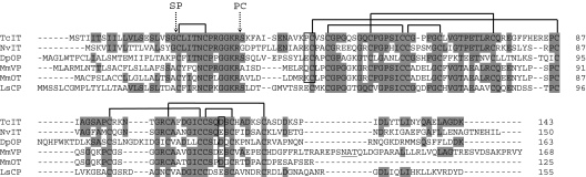
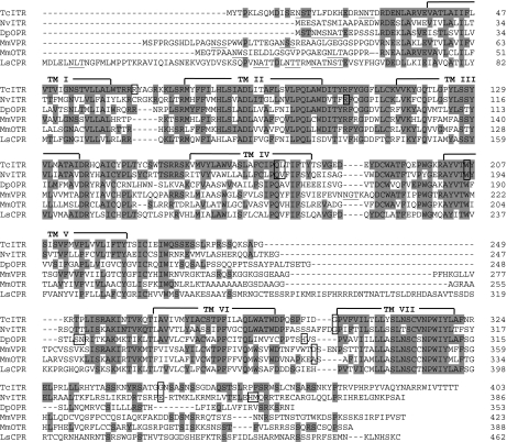
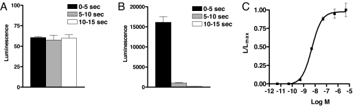
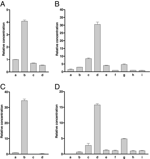
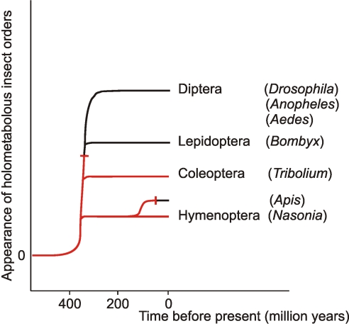
More than 20 years ago, an oxytocin/vasopressin-like peptide, CLITNCPRGamide, was isolated from the locust, Locusta migratoria [Proux JP, et al. (1987) Identification of an arginine vasopressin-like diuretic hormone from Locusta migratoria. Biochem Biophys Res Commun 149:180-186]. However, no similar peptide could be identified in other insects, nor could its prohormone be cloned, or its physiological actions be established. Here, we report that the recently sequenced genome from the red flour beetle Tribolium castaneum contains a gene coding for an oxytocin/vasopressin-like peptide, identical to the locust peptide, which we named inotocin (for insect oxytocin/vasopressin-like peptide) and a gene coding for an inotocin G protein-coupled receptor (GPCR). We cloned the Tribolium inotocin preprohormone and the inotocin GPCR and expressed the GPCR in CHO cells. This GPCR is strongly activated by low concentrations of inotocin (EC(50), 5 x 10(-9) M), demonstrating that it is the inotocin receptor. Quantitative RT-PCR (qPCR) showed that in adult Tribolium, the receptor is mainly expressed in the head and much less in the hindgut and Malpighian tubules, suggesting that the inotocin/receptor couple does not play a role in water homeostasis. Surprisingly, qPCR also showed that the receptor is 30x more expressed in the first larval stages than in adult animals. The inotocin/receptor couple can also be found in the recently sequenced genome from the parasitic wasp Nasonia vitripennis but not in any other holometabolous insect with a completely sequenced genome (12 Drosophila species, the malaria mosquito Anopheles gambiae, the yellow fever mosquito Aedes aegypti, the silk worm Bombyx mori, and the honey bee Apis mellifera), suggesting that this neuropeptide system is confined to basal holometabolous insects. Furthermore, we identified an oxytocin/vasopressin-like peptide and receptor in the recently sequenced genome from the water flea Daphnia pulex (Crustacea). To our knowledge, this is the first report on the molecular cloning of an oxytocin/vasopressin-like receptor and its ligand from arthropods.


Conflict of interest statement

The authors declare no conflict of interest.

Figures

Fig. 1.
Fig. 1.
Alignment of the preprohormones for T. castaneum inotocin (TcIT; GenBank accession no. EU156489), N. vitripennis inotocin (NvIT; GenBank accession no. XP_001606547), D. pulex oxytocin/vasopressin-like peptide (DpOP), mouse (Mus musculus) vasopressin (MmVP; GenBank accession no. NP_033862), mouse oxytocin (MmOT; GenBank accession no. NP_035155), and L. stagnalis conopressin (LsCP; GenBank accession no. AAA29289). The amino acid residues identical to TcIT are highlighted. The common intron positions are indicated by vertical boxes. The cleavage sites for the signal peptidases (SP) and prohormone convertases (PC) are indicated by dotted arrows. These proteinases liberate dodecapeptides that are further processed to the amidated oxytocin/vasopressin-like nonapeptides. There are three domains containing cystine bridges: the nonapeptide domain (one cystine bridge), and two neurophysin domains containing four and three cystine bridges, respectively. This overall structure and many amino acid sequences, have been conserved in all six proteins. The N. vitripennis and D. pulex sequences have been annotated, the others have been experimentally determined (cDNA cloning). The introns for the L. stagnalis sequence could not be determined because of the lack of genomic information.
Fig. 2.
Fig. 2.
Alignment of the T. castaneum inotocin receptor (TcITR; GenBank accession no. EU128495), N. vitripennis inotocin-like receptor (NvITR; GenBank accession no. XP_001600203), D. pulex oxytocin/vasopressin-like receptor (DpOPR), mouse V1a vasopressin receptor (MmVPR; GenBank accession no. NP_058543), mouse oxytocin receptor (MmOTR; GenBank accession no. NP_001074616), and L. stagnalis conopressin receptor (LsCPR; GenBank accession no. AAA91998) proteins. The residues identical to TcITR are highlighted. The transmembrane α-helices are indicated by TMI-TMVII. Introns are indicated by boxes. Potential glycosylation sites, occurring mainly in the extracellular N terminus, are underlined. The N. vitripennis and D. pulex sequences have been annotated, the others have been experimentally determined (cDNA cloning). The introns for the L. stagnalis sequence could not be determined because of the lack of genomic information.
Fig. 3.
Fig. 3.
Bioluminescence responses of non-transfected CHO/G-16 cells (A) and CHO/G-16 cells, expressing the inotocin receptor gene (B), 0–5 s (black), 5–10 s (gray) and 10–15 s (white) after addition of 5 × 10⁻⁶ M inotocin. Note that the scales in A and B are different. (C) Dose–response curve of the effect of inotocin on CHO/G-16 cells expressing the inotocin receptor. The EC50 of inotocin is 5 × 10⁻⁹ M. SEM are given as vertical bars, which are sometimes smaller than the symbols used (squares or lines). In these cases, only the symbols are given. In addition to inotocin, the inotocin receptor is also activated by Arg- and Lys-conopressins, vasotocin, oxytocin, and isotocin (EC50 values, >10−6 M). Vasopressin did not activate the receptor. Thirty-three other insect neuropeptides and eight biogenic amines (SI Text, Materials and Methods) did also not activate the receptor (tested up to 10−5 M).
Fig. 4.
Fig. 4.
qPCR of inotocin receptor and preprohormone mRNA in adults or different developmental stages from Tribolium. In each column, at least 60 animals were pooled. The qPCR experiments were run as triplets; the bars (which sometimes are smaller than the lines) represent SEM. Two different ribosomal proteins (rpL32 and rps3) were used as references and gave similar results. Each experiment was repeated at least twice. (A) Inotocin receptor mRNA in adult mixed male and female animals: a, whole bodies; b, heads; c, torsi (body minus head); and d, pooled hindguts and Malpighian tubules. The receptor mRNA concentrations given are relative to bar a (=1). (B) Inotocin receptor mRNA in different developmental stages (the sexes were mixed except for adult animals): a, eggs 0–24 h after egg laying; b, eggs 24–48 h after egg laying; c, eggs 48–72 h after egg laying; d, larvae 96–120 h after egg laying (≈0–1 d after hatching); e, larvae 15–16 d after egg laying; f, larvae 20–21 d after egg laying; g, pupae (24–25 d after egg laying); h, adult female, and i, adult male animals (27 d or more after egg laying). The receptor mRNA concentrations given are relative to bar h (=1). (C) Inotocin preprohormone mRNA in the same tissues as given in A. The mRNA concentrations given are relative to bar a (=1). (D) Inotocin preprohormone mRNA in the same developmental stages as given in B. The mRNA concentrations given are relative to bar h (=1).
Fig. 5.
Fig. 5.
Schematic representation of the appearance of the major orders of holometabolous insects and the occurrence of the inotocin hormonal system (highlighted in red). This hormonal system has been conserved only in the evolutionary lines leading to basal holometabolous insects: Coleoptera (beetles) and Hymenoptera (wasps). The inotocin system must have been abandoned at least two times during the evolution of the Holometabola (see dead-end signs).

References

    1. Acher R, Chauvet J. La structure de la vasopressin de boeuf. Biochim Biophys Acta. 1953;12:487–488. - PubMed
    1. du Vigneaud V, Ressler C, Trippett S. The sequence of amino acids in oxytocin, with a proposal for the structure of oxytocin. J Biol Chem. 1953;205:949–957. - PubMed
    1. du Vigneaud V, Lawler HC, Popenoe EA. Enzymatic cleavage of glycinamide from vasopressin and a proposed structure for this pressor-antidiuretic hormone of the posterior pituitary. J Am Chem Soc. 1953;75:4880–4881.
    1. Schmale H, Heinsohn S, Richter D. Structural organization of the rat gene for the arginine vasopressin-neurophysin precursor. EMBO J. 1983;2:763–767. - PMC - PubMed
    1. Ivell R, Schmale H, Richter D. Vasopressin and oxytocin precusors as model preprohormones. Neuroendocrinology. 1983;37:235–240. - PubMed

Publication types

Associated data

LinkOut - more resources