

Human muscle gene expression following resistance exercise and blood flow restriction
- PMID: 18317375
- PMCID: PMC5088719
- DOI: 10.1249/MSS.0b013e318160ff84
Human muscle gene expression following resistance exercise and blood flow restriction
Erratum in
- Med Sci Sports Exerc 2008 Jun;40(6):1191.. Takashi, Abe [corrected to Abe, Takashi].
Abstract
Introduction: Blood flow restriction in combination with low-intensity resistance exercise (REFR) increases skeletal muscle size to a similar extent as compared with traditional high-intensity resistance exercise training. However, there are limited data describing the molecular adaptations that occur after REFR.
Purpose: To determine whether hypoxia inducible factor-1 alpha (HIF-1alpha) and REDD1 mRNA are expressed differently in REFR compared with low-intensity resistance exercise with no blood flow restriction (CONTROL). Secondly, to determine whether low-intensity resistance exercise is able to induce changes in mRNA expression of several anabolic and catabolic genes as typically seen with high-intensity resistance exercise.
Methods: Six subjects were studied at baseline and 3 h after a bout of leg resistance exercise (20% 1RM) in REFR and CONTROL subjects. Each subject participated in both groups, with 3 wk separating each visit. Muscle biopsy samples were analyzed for mRNA expression, using qRT-PCR.
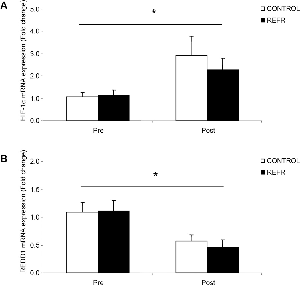
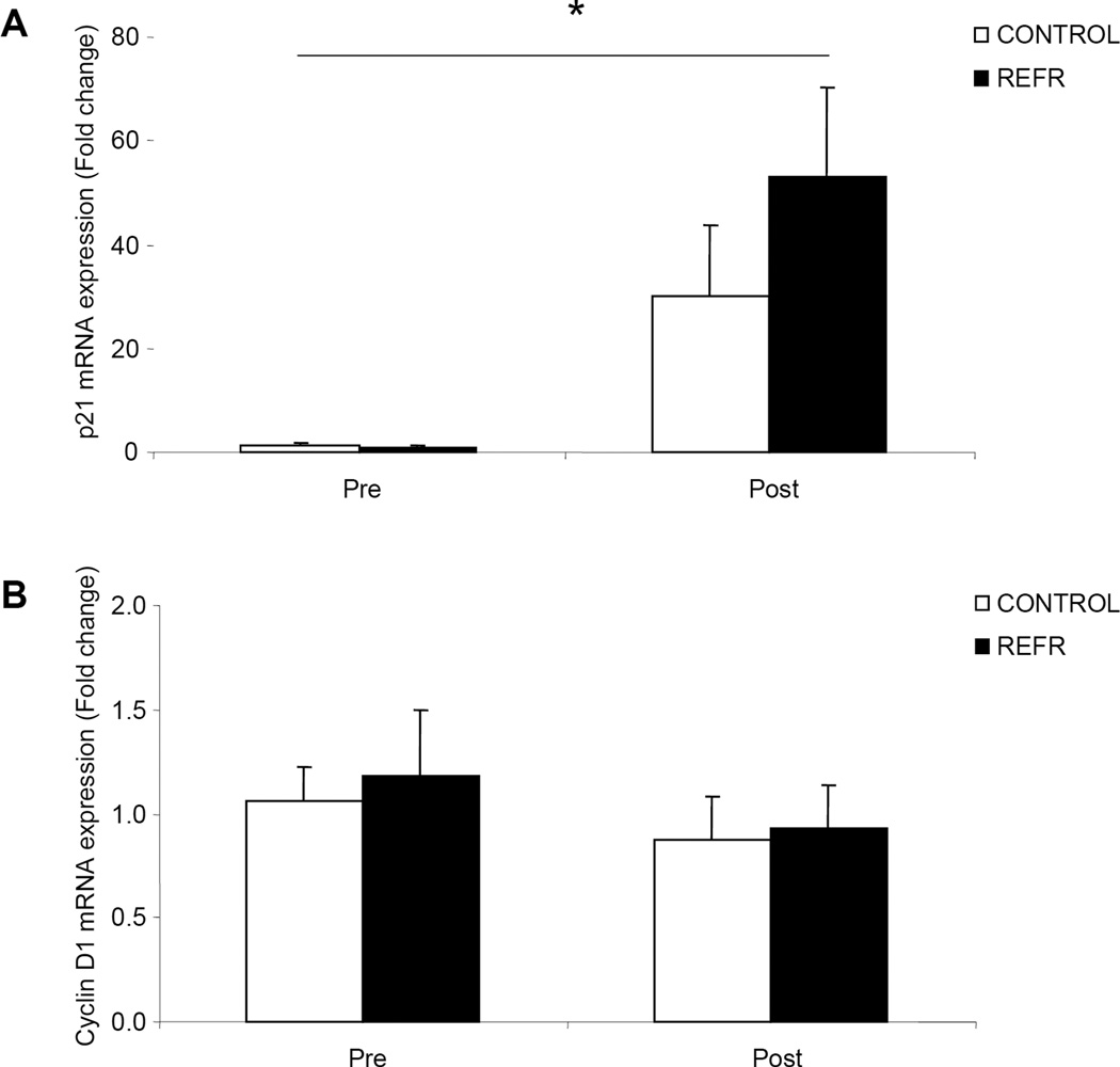
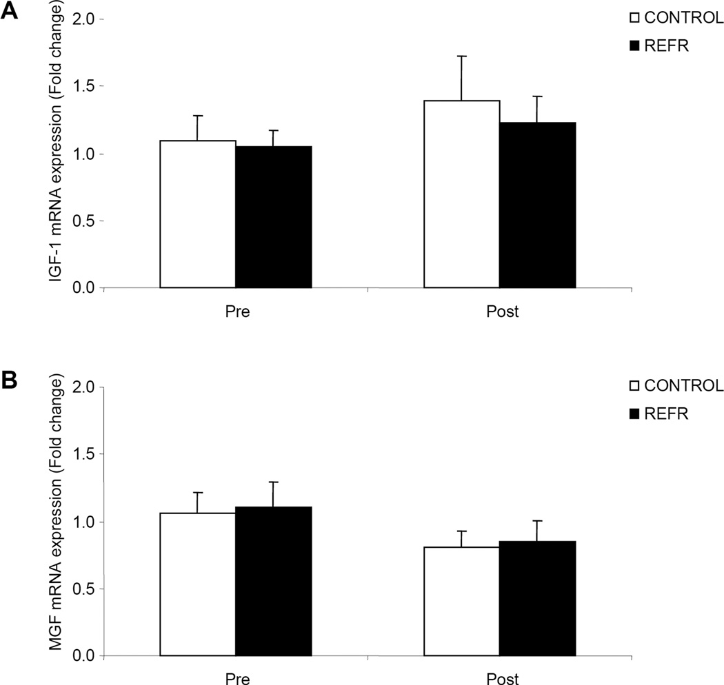
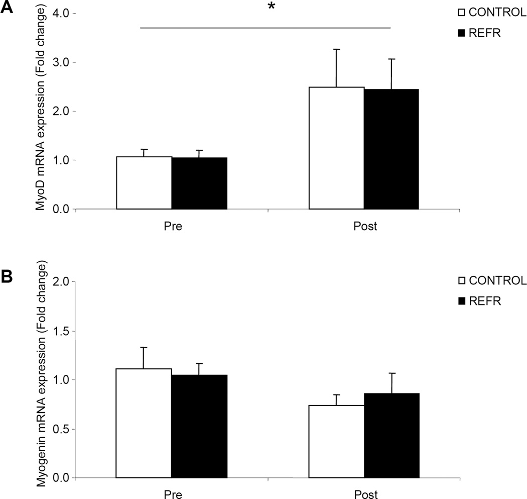
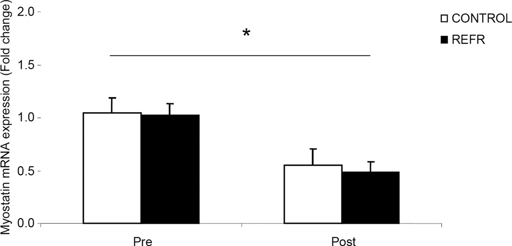
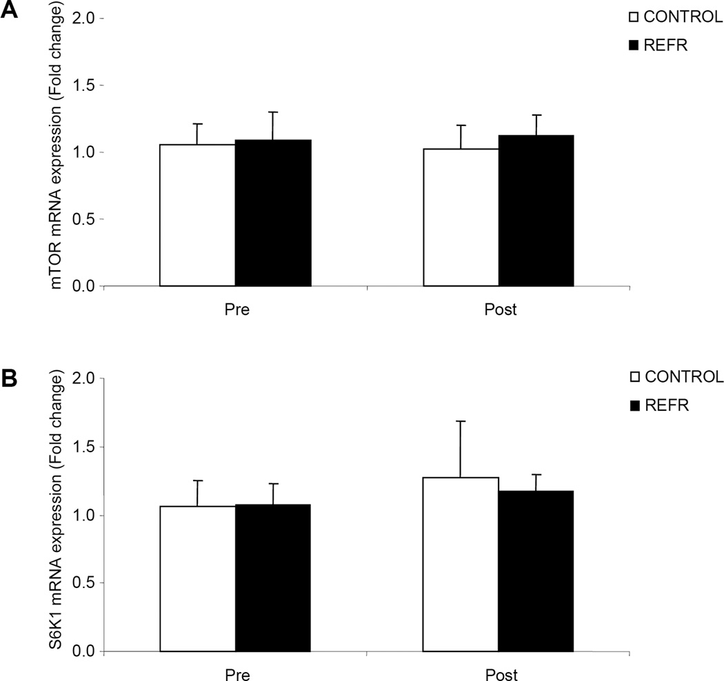
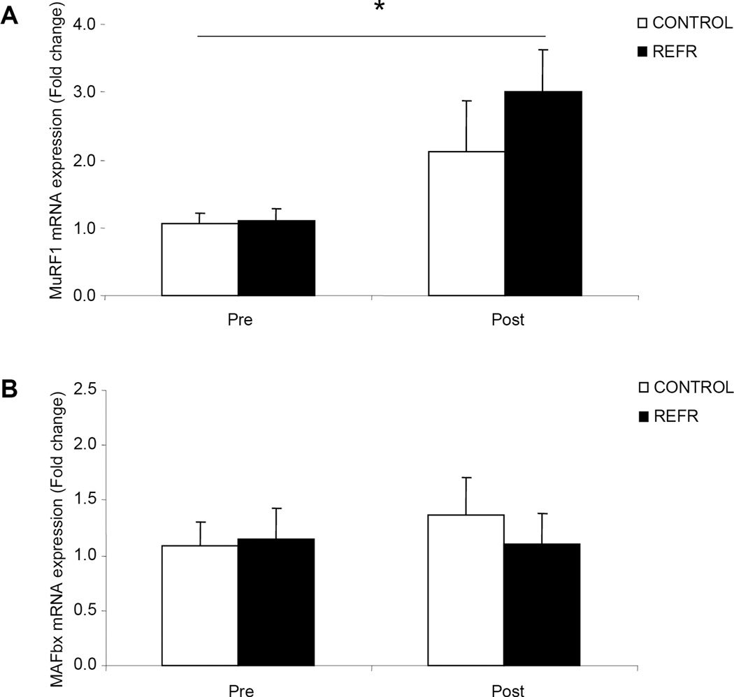
Result: Our primary finding was that there were no differences between CONTROL and REFR for any of the selected genes at 3 h after exercise (P > 0.05). However, low-intensity resistance exercise increased HIF-1alpha, p21, MyoD, and muscle RING finger 1 (MuRF1) mRNA expression and decreased REDD1 and myostatin mRNA expression in both groups (P < 0.05).
Conclusion: Low-intensity resistance exercise can alter skeletal muscle mRNA expression of several genes associated with muscle growth and remodeling, such as REDD1, HIF-1alpha, MyoD, MuRF1, and myostatin. Further, the results from REFR and CONTROL were similar, indicating that the changes in early postexercise gene expression were attributable to the low-intensity resistance exercise bout, and not blood flow restriction.
Conflict of interest statement
The authors report no conflict of interest or endorsement by ACSM.
Figures
References
-
- Abe T, Kearns CF, Sato Y. Muscle size and strength are increased following walk training with restricted venous blood flow from the leg muscle, Kaatsu-walk training. J Appl Physiol. 2006;100(5):1460–1466. - PubMed
-
- Abe T, Yasuda T, Midorikawa T, Sato Y, Kearns CF, Inoue K, Koizumi K, Ishii N. Skeletal muscle size and circulating IGF-1 are increased after two weeks of twice daily KAATSU resistance training. Int J Kaatsu Training Res. 2005;1(6–12)
-
- Ameln H, Gustafsson T, Sundberg CJ, Okamoto K, Jansson E, Poellinger L, Makino Y. Physiological activation of hypoxia inducible factor-1 in human skeletal muscle. Faseb J. 2005;19(8):1009–1011. - PubMed
-
- Arsham AM, Howell JJ, Simon MC. A novel hypoxia-inducible factor-independent hypoxic response regulating mammalian target of rapamycin and its targets. J Biol Chem. 2003;278(32):29655–29660. - PubMed
-
- Bamman MM, Shipp JR, Jiang J, Gower BA, Hunter GR, Goodman A, McLafferty CL, Jr, Urban RJ. Mechanical load increases muscle IGF-I and androgen receptor mRNA concentrations in humans. Am J Physiol Endocrinol Metab. 2001;280(3):E383–E390. - PubMed
Publication types
MeSH terms
Substances
Grants and funding
LinkOut - more resources
Full Text Sources
Medical
Miscellaneous
