. 2008 Apr;118(4):1468-78.
doi: 10.1172/JCI33869.

Neutrophil gelatinase-associated lipocalin mediates 13-cis retinoic acid-induced apoptosis of human sebaceous gland cells

Affiliations

Neutrophil gelatinase-associated lipocalin mediates 13-cis retinoic acid-induced apoptosis of human sebaceous gland cells

Amanda M Nelson et al. J Clin Invest. 2008 Apr.

Abstract

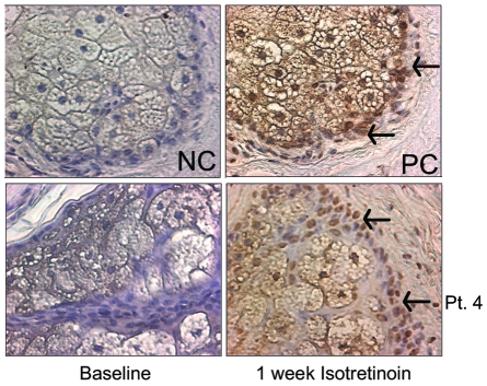
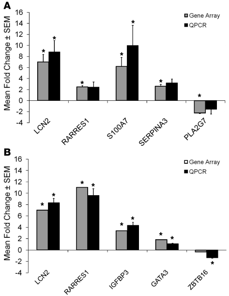
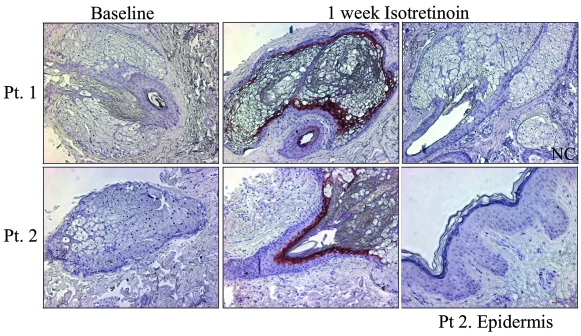
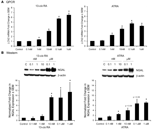
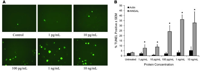
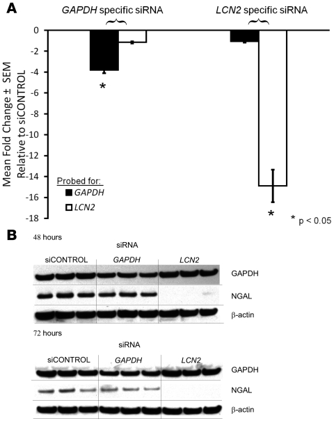
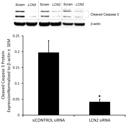
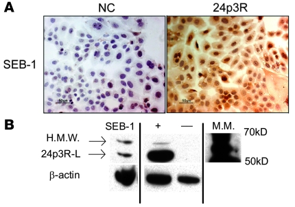
13-cis retinoic acid (13-cis RA; also known as isotretinoin) is the most potent agent available for treatment of acne. It is known that the drug induces apoptosis in cells cultured from human sebaceous glands, but its mechanism of action has not been determined. In this study, skin biopsies were taken from 7 patients with acne prior to and at 1 week of treatment with 13-cis RA. TUNEL staining confirmed that 13-cis RA induced apoptosis in sebaceous glands. Transcriptional profiling of patient skin and cultured human sebaceous gland cells (SEB-1 sebocytes) indicated that lipocalin 2 was among the genes most highly upregulated by 13-cis RA. Lipocalin 2 encodes neutrophil gelatinase-associated lipocalin (NGAL), which functions in innate immune defense and induces apoptosis of murine B lymphocytes. Increased immunolocalization of NGAL was noted in patients' sebaceous glands following treatment with 13-cis RA, and recombinant NGAL induced apoptosis in SEB-1 sebocytes. Furthermore, apoptosis in response to 13-cis RA was inhibited in the presence of siRNA to lipocalin 2. These data indicate that NGAL mediates the apoptotic effect of 13-cis RA and suggest that agents that selectively induce NGAL expression in sebaceous glands might represent therapeutic alternatives to the use of 13-cis RA to treat individuals with acne.


Figures

Figure 1
Figure 1. TUNEL staining is increased in patients’ sebaceous glands at 1 week of isotretinoin treatment.
Skin sections were obtained from paraffin blocks of 7 patients sampled at baseline and 1 week of isotretinoin and were subjected to TUNEL-peroxidase assay, followed by counterstaining with hematoxylin. At least 2 skin sections from each time point were analyzed from every patient. Patient 7 was omitted from the analysis as no sebaceous glands were found in baseline biopsy sections. Results were quantified by counting TUNEL-positive cells / total cells in sebaceous glands. Representative sections from Patient 4 (Pt. 4) taken at baseline and at 1 week of isotretinoin treatment are shown. TUNEL staining was strongest in the nuclei of sebocytes in the basal layer of the sebaceous gland (arrows) and in early differentiated sebocytes adjacent to the basal layer. Apoptosis was selective for the sebaceous gland, as no apoptosis was detected within the epidermis (not shown). Negative control (NC) consists of sections from a skin specimen treated with DNase I and processed without terminal transferase enzyme. Positive control (PC) consists of sections from a skin specimen treated with DNase I and subjected to the full TUNEL assay. Original magnification, ×400.
Figure 2
Figure 2. QPCR verification of gene array changes.
In order to verify the direction and magnitude of the changes in gene expression induced by 13-cis RA in the gene microarrays, QPCR was performed using primers to select genes whose expression was significantly changed by 13-cis RA in the array analysis. (A) Comparison of array analysis and QPCR on RNA obtained from patient skin biopsies at baseline and 1 week of 13-cis RA treatment. Data represent the mean ± SEM of the fold change in gene expression as determined by QPCR in 5 subjects compared to array analysis performed in 6 subjects. RARRES1, retinoic acid receptor responder 1; S100A7, psoriasin; SERPINA3, serine proteinase inhibitor A3; PLA2G7, phospholipase A2 group 7. (B) Comparison of array analysis and QPCR on RNA obtained from SEB-1 sebocytes incubated for 72 hours in the presence or absence of 13-cis RA. Data represent the mean ± SEM of the fold change in gene expression as determined by QPCR in 8 samples compared to array analysis performed in 3 samples. QPCR results were analyzed by REST-XL software program and *P < 0.05 was considered significant. IGFBP3, insulin-like growth factor–binding protein 3; GATA3, GATA-binding protein 3; ZBTB16, zinc finger and BTB domain-containing 16.
Figure 3
Figure 3. NGAL expression is increased in sebaceous glands in patients biopsied at 1 week of isotretinoin treatment.
Immunohistochemistry using an antibody to NGAL was performed on skin sections taken at baseline and at 1 week of isotretinoin treatment. Sections were incubated overnight with a 1:50 dilution of mouse monoclonal LCN2/NGAL antibody and developed using AEC chromagen (red). All sections were counterstained with hematoxylin. Representative images at baseline and after 1 week isotretinoin from Patients 1 and 2 are shown. An image of the epidermis after isotretinoin treatment from Patient 2 is shown. NGAL was expressed in the sebaceous gland and duct of samples of skin taken at 1 week of isotretinoin therapy. NGAL was not expressed in the epidermis. The amount of NGAL staining varies among patients and individual patient results are shown in Table 4. Negative control consists of normal human skin incubated with normal mouse IgG1 antibody. Original magnification, ×100.
Figure 4
Figure 4. 13-cis RA and ATRA increase LCN2 mRNA and NGAL protein expression in SEB-1 sebocytes.
SEB-1 sebocytes were treated with vehicle as a control (C), 13-cis RA (0.1 nM, 1 nM, 10 nM, 0.1 μM, or 1 μM) or ATRA (0.1 nM, 1 nM, 10 nM, 0.1 μM, or 1 μM) for 48 and 72 hours. (A) LCN2 mRNA expression was verified by QPCR after 48 hours of retinoid treatment. Data represent mean ± SEM of the fold change in gene expression as determined by QPCR of 5 independent samples. Statistical analysis was performed with REST-XL software program and considered significant if *P < 0.05. (B) Protein expression was verified by western blot at 72 hours of retinoid treatment. Blots were incubated with primary antibody to NGAL as well as β-actin as a loading control. Blots were analyzed by densitometry and normalized to β-actin. The graph represents normalized fold-change values (mean ± SEM) relative to control for a minimum of 3 independent blots. Statistical analysis was performed with paired t test; *P < 0.05.
Figure 5
Figure 5. NGAL increases TUNEL staining in SEB-1 sebocytes.
SEB-1 sebocytes were treated with vehicle as a control, 1 pg/ml, 10 pg/ml, 100 pg/ml, 1 ng/ml, or 10 ng/ml of purified rhNGAL protein (R&D Systems) or the same concentrations of human actin protein for 24 hours. (A) Representative images of the TUNEL assay using rhNGAL are shown. Original magnification, ×200. (B) Quantification of the percentage of TUNEL-positive stained cells per treatment at 24 hours. Data represent mean ± SEM; n = 4–8. Parallel experiments to control for nonspecific effects of protein were performed using human actin protein (n = 2). The percentage of TUNEL-positive cells is less than 5% with all concentrations of actin, which is similar to control values. rhNGAL significantly increased TUNEL staining compared to control over a wide range of concentrations with maximal induction noted at 1 ng/ml. Statistical analyses were performed between vehicle control and each treatment concentration of rhNGAL using 2-factor ANOVA with replication; *P < 0.05.
Figure 6
Figure 6. siRNA to LCN2 specifically knocks down LCN2 expression in SEB-1 sebocytes in the presence of 13-cis RA.
SEB-1 sebocytes (2 × 10⁶ cells per 100 μl reaction mixture) were nucleofected with 1 μg siCONTROL, GAPDH (as a specificity control), or LCN2 siRNA. 13-cis RA (0.1 μM) was added 24 hours after nucleofection, and cells were incubated for 48 and 72 hours. Extent of siRNA knockdown of gene expression was verified by QPCR and western blotting for LCN2 and GAPDH after 13-cis RA treatment. GAPDH and LCN2 gene expression were successfully inhibited in their respective samples. (A) QPCR analysis of LCN2 and GAPDH mRNA levels at 48 hours of 0.1 μM 13-cis RA treatment. The expression of LCN2 mRNA was successfully decreased 15-fold by the LCN2 siRNA compared with siCONTROL, whereas expression of GAPDH was minimally affected by siRNA to LCN2. GAPDH mRNA expression was decreased 4-fold by the specific GAPDH siRNA when compared with siCONTROL, whereas siRNA to GAPDH had minimal effects on expression of mRNA for LCN2. Data represent mean ± SEM; n = 6. *P < 0.05 as determined by REST-XL program. (B) Western analysis of GAPDH and NGAL protein levels. NGAL protein expression is decreased to undetectable levels by western blotting at 48 and 72 hours of 0.1 μM 13-cis RA treatment with LCN2 siRNA. Representative blots of 4 independent experiments are shown.
Figure 7
Figure 7. siRNA to NGAL blocks the induction of cleaved caspase 3 by 13-cis RA in SEB-1 sebocytes.
Under siRNA conditions described in Figure 6, immunoblotting revealed that siRNA to LCN2 decreases expression of cleaved caspase 3 by approximately 5-fold compared with siRNA control. Blots were incubated with primary antibodies to cleaved caspase 3 as well as β-actin as a loading control to normalize densitometry values. Three independent scrambled siCONTROL (SCRAM) and LCN2 western blots are shown. p17 and p19 are the cleaved or active fragments of caspase 3. Graph represents the normalized expression of cleaved caspase 3 from 5 independent western blots. These data indicate that NGAL mediates the apoptotic response of SEB-1 sebocytes to 13-cis RA. Statistical analysis was performed with paired t test, mean ± SEM; *P < 0.05.
Figure 8
Figure 8. SEB-1 sebocytes express the receptor for NGAL.
(A) SEB-1 sebocytes were grown under standard conditions and immunocytochemistry was performed using an antibody to the murine 24p3R. Slides were counterstained with hematoxylin. Negative control (NC) was processed with normal rabbit IgG antibody in place of the primary antibody. Scale bar: 50 μm. Immunoreactivity for the 24p3R localizes to the cytoplasm of SEB-1 sebocytes. (B) Western analysis confirms presence of the receptor and indicates 2 receptor isoforms are present in SEB-1 sebocytes: high molecular weight (H.M.W.) and 24p3R long (24p3R-L). Positive ([+]; HEK 293 cell lysate) and negative ([–]; T47D cell lysate) controls provided by are shown. All samples were run on the same gel but were noncontiguous. Blot shown is representative of 3 independent experiments.

References

    1. Reynolds C.P., Matthay K.K., Villablanca J.G., Maurer B.J. Retinoid therapy of high-risk neuroblastoma. Cancer Lett. 2003;197:185–192. doi: 10.1016/S0304-3835(03)00108-3. - DOI - PubMed
    1. Landthaler M., Kummermehr J., Wagner A., Plewig G. Inhibitory effects of 13-cis retinoic acid on human sebaceous glands. . Arch. Dermatol. Res. 1980;269:297–309. doi: 10.1007/BF00406424. - DOI - PubMed
    1. Strauss J.S., Stranieri A.M., Farrell L.N., Downing D.T. The effect of marked inhibition of sebum production with 13-cis retinoic acid on skin surface lipid composition. . J. Invest. Dermatol. 1980;74:66–67. doi: 10.1111/1523-1747.ep12519851. - DOI - PubMed
    1. Goldstein J.A., Comite H., Mescon H., Pochi P.E. Isotretinoin in the treatment of acne. Arch. Dermatol. 1982;118:555–558. doi: 10.1001/archderm.118.8.555. - DOI - PubMed
    1. Peck G.L. Prolonged remissions of cystic acne with 13-cis retinoic acid. . N. Engl. J. Med. 1979;300:329. - PubMed

Publication types

MeSH terms