. 2008 Apr;118(4):1437-49.
doi: 10.1172/JCI32638.

Targeted mutation of mouse skeletal muscle sodium channel produces myotonia and potassium-sensitive weakness

Affiliations

Targeted mutation of mouse skeletal muscle sodium channel produces myotonia and potassium-sensitive weakness

Lawrence J Hayward et al. J Clin Invest. 2008 Apr.

Abstract

Hyperkalemic periodic paralysis (HyperKPP) produces myotonia and attacks of muscle weakness triggered by rest after exercise or by K+ ingestion. We introduced a missense substitution corresponding to a human familial HyperKPP mutation (Met1592Val) into the mouse gene encoding the skeletal muscle voltage-gated Na+ channel NaV1.4. Mice heterozygous for this mutation exhibited prominent myotonia at rest and muscle fiber-type switching to a more oxidative phenotype compared with controls. Isolated mutant extensor digitorum longus muscles were abnormally sensitive to the Na+/K+ pump inhibitor ouabain and exhibited age-dependent changes, including delayed relaxation and altered generation of tetanic force. Moreover, rapid and sustained weakness of isolated mutant muscles was induced when the extracellular K+ concentration was increased from 4 mM to 10 mM, a level observed in the muscle interstitium of humans during exercise. Mutant muscle recovered from stimulation-induced fatigue more slowly than did control muscle, and the extent of recovery was decreased in the presence of high extracellular K+ levels. These findings demonstrate that expression of the Met1592ValNa+ channel in mouse muscle is sufficient to produce important features of HyperKPP, including myotonia, K+-sensitive paralysis, and susceptibility to delayed weakness during recovery from fatigue.


Figures

Figure 1
Figure 1. Knock-in of human Met1592Val variant into mouse NaV1.4 gene.
(A) Na+ channel topology and the location of the Met1592Val substitution within the S6 segment of domain IV (top). The mNaV1.4 targeting sequence (bottom) encompassed exons 17–24 (black boxes) and the 3′-UTR of exon 24 (hatched area), while the PGKneo selection gene was inserted within intron 23. The Met→Val mutation (*) and the silent HpaI site (H) are indicated within exon 24. Bold X’s indicate the limits of the targeting sequence. Shown are the internal and external hybridization probe regions and restriction enzyme sites for AvrII (A), AflII (A2), KpnI (K), NheI (N), EcoRI (R), SacI (S), and ScaI (Sc). (B) Southern blot of NheI-digested mouse genomic DNA (10 μg per lane) hybridized with the internal 3′-UTR probe demonstrates a 4.5-kb wild-type allele or a 6.1-kb allele produced by insertion of the PGKneo gene. Genotype is indicated for wild-type (+/+), heterozygous mutant (+/m), and homozygous mutant (m/m) alleles. (C) Southern blot of DNA digested with AflII plus HpaI plus ScaI and hybridized to the external probe identifies an 8.0-kb wild-type allele (ScaI–AflII fragment) and a 4.4 kb mutant allele (HpaI–AflII fragment). (D) Genotyping by analysis of a 1.39-kb PCR product amplified from exon 24 using primers a and b (see Supplemental Methods). The mutant allele contained a novel HpaI site and lacked one of the 5 NspI sites present in the wild-type allele (indicated by thick vertical lines under the asterisk, with sequence shown below). The lane labeled “M” shows a 100-bp marker DNA ladder.
Figure 2
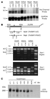
Figure 2. Mutant Na+ channel mRNA and protein are expressed in mouse skeletal muscle tissue.
(A) Northern blots of total RNA extracted from pooled hind-limb skeletal muscle, quadriceps, gastrocnemius/soleus, heart, or brain tissues (5 μg loaded per lane; n = 3–8 mice per group). The 3′-UTR probe specific for mNaV1.4 hybridized to an 8.5-kb full-length Na+ channel mRNA in both normal (+/+) and mutant (+/m) muscle samples but not those from heart or brain (top panel). The blot was rehybridized with a GAPDH probe (bottom panel). (B) RT-PCR of total RNA from (+/+) or (+/m) mouse muscle using primers c (exon 23) and b (exon 24). Digestion of the mutant RT-PCR product with HpaI (producing 0.89-kb and 0.54-kb fragments) or NspI (preserving a 0.71-kb fragment) demonstrated that approximately 30%–50% of transcripts in mutant muscle encoded the Met1592Val variant. The lane labeled “M” shows a 100-bp marker DNA ladder. (C) Western blot of membrane proteins isolated from (+/+), (+/m), or (m/m) mouse quadriceps muscle, heart (HT), and brain (BN) tissues (10 μg loaded per lane). The L/D3 monoclonal antibody specifically recognizes NaV1.4 but not cardiac or brain Na+ channel isoforms.
Figure 3
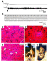
Figure 3. Heterozygous mutant (+/m) muscle from young mice exhibited myotonia and only subtle myopathy, while homozygous (m/m) mutant muscle exhibited abnormal limb clasping and prominent myopathic changes.
(A) Electromyography of hind-limb muscle from normal (+/+) anesthetized mice showed electrical silence except upon insertion (arrow) or movement of the recording electrode. Electromyograms from mutant (+/m) mice under the same conditions, without exception, exhibited high background activity and audible characteristics consistent with widespread myotonia (n > 60 for both groups). (B) Runs of myotonia (sustained repetitive firing with varying amplitude and frequency) occurred spontaneously or could be easily elicited upon needle movement for (+/m) muscle (shown) but not for normal muscle (see Supplemental Videos 1 and 2). (CE) H&E staining of hamstring muscle from a (+/+) mouse at 4 months of age exhibited a normal pattern of fiber size variation and peripheral nuclei (C), while that from a (+/m) mouse at 4 months showed mildly increased internalized nuclei (D). The same muscle from a (m/m) mouse at 2.8 months exhibited increased fiber size variation, frequent internalized nuclei, and large scattered vacuoles at various stages of evolution (E). Scale bars in CE: 100 μm. (F) Normal extension of hind limbs observed for a (+/m) mouse at 2.8 months of age upon being held by the tail. (G) Abnormal clasping response exhibited by a (m/m) sibling at 2.8 months. This (m/m) mouse also had prominent hind-limb weakness and decreased locomotor activity compared with its (+/m) sibling (see Supplemental Video 4).
Figure 4
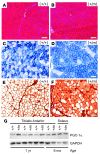
Figure 4. Mutant (+/m) muscles from older mice exhibited mild myopathic changes, a more oxidative fiber type, and upregulation of the transcriptional coactivator PGC-1α.
(A and B) H&E staining of tibialis anterior muscle from a normal (+/+) mouse at 24 months of age exhibited a normal pattern of fiber size variation and peripheral nuclei (A), while that from a mutant (+/m) sibling mouse showed increased fiber size variation and more frequent internalized nuclei (B). (C and D) Succinate dehydrogenase (SDH) staining of serial sections revealed a mixed pattern of oxidative (dark) and glycolytic (light) fibers for tibialis anterior muscle from the normal (+/+) mouse (C), while that from the (+/m) mouse showed an increase of SDH-positive fibers, indicating a more oxidative phenotype (D). (E and F) Immunostaining of serial sections with A4.74 myosin antibody confirmed a large increase in fast oxidative fibers (type IIA) in the mutant (+/m) compared with the normal (+/+) muscle. Scale bars in AF: 100 μm. (G) Western blot (2 μg of soluble muscle lysate protein per lane as determined by BCA assay) showed increased expression of PGC-1α by 2.1 ± 0.5–fold in tibialis anterior muscle from 1-year-old (+/m) mice compared with controls.
Figure 5
Figure 5. Reduced force and slowed relaxation of mutant (+/m) compared with normal (+/+) EDL muscle contributed to altered contractile properties.
(A) Single-twitch contraction produced by a 1-ms supramaximal current pulse to isolated EDL muscle from a mutant (+/m) or a normal (+/+) mouse. Responses were normalized to the peak force for comparison of kinetics. Bar graphs (mean ± SD) indicate decreased peak force by 2.3-fold, no change for time to peak, and increased T1/2R by 1.7-fold for mutant (n = 9, 10.6 ± 2.2 months old) compared with normal (n = 5, 11.4 ± 1.8 months old) EDL muscles. (B) Tetanic contraction produced by 1-ms pulse trains lasting 0.5 seconds at 20, 50, or 100 Hz for the same EDL muscles as in A. Records are normalized to the peak force during 100-Hz stimulation. The kinetics of force buildup (tetanic rise τ) and the tetanic T1/2R from the last stimulus were both prolonged for mutant compared with normal EDL during 50- and 100-Hz stimulation. The ratio of maximal tetanic force to single-twitch force was significantly increased for mutant compared with control muscle at 50- and 100-Hz stimulation. Bar graphs show mean ± SD. *P < 0.01, 2-tailed Student’s t test.
Figure 6
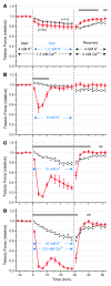
Figure 6. Raising [K+]o to 10 mM produced sustained weakness that was greater for mutant (+/m) compared with normal (+/+) EDL muscle.
Isolated EDL muscles from 8.8 ± 0.3–month-old mutant mice or sibling controls were equilibrated for more than 30 minutes at 25°C in starting bath solution containing 4 mM [K+] and 1.3 mM [Ca2+]. Tetanic stimuli (0.5-ms, 70-mA current pulses for 300 ms at 125 Hz) were applied every 3 minutes, and the peak tetanic responses (mean ± SEM, n = 5–6 for each group) are shown normalized to the starting values, 15.7 ± 2.3 g for (+/m) and 18.4 ± 0.6 g for (+/+) muscle. Following each test condition, the bath was replaced with a recovery solution identical to the starting solution except containing approximately 3-fold higher [Ca2+] (4 mM) to help repolarize the membrane (37). (A) Lowering [K+]o to 1.2 mM produced only mild force reduction that did not differ between mutant and control muscles. (B) Raising [K+]o to 8 mM produced only transient weakness for mutant muscle, followed by recovery of force within 15 minutes. (C) Raising [K+]o to 10 mM caused pronounced and sustained weakness for mutant but not control muscle that was fully reversible in the recovery solution. (D) Lowering [Ca2+]o to 0.5 mM exacerbated the weakness produced by elevated [K+]o. Gray bars indicate significant differences (P < 0.05 by repeated-measures ANOVA with Bonferroni correction) between mutant (red circles) and control (open squares) responses.
Figure 7
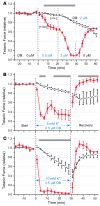
Figure 7. Mutant (+/m) EDL was more sensitive than control (+/+) muscle to inhibition of force by ouabain (OB), which also exacerbated the weakness produced by elevated [K+]o.
Isolated EDL muscles from 8.9 ± 0.2–month-old mutant mice or sibling controls were equilibrated in bath containing 4 mM [K+]o and 1.3 mM [Ca2+]o and stimulated as in Figure 6; normalized peak tetanic responses (mean ± SEM) are shown. (A) Mutant EDL was highly sensitive to 0.5–2.0 μM ouabain, which affected control muscle much more slowly. (B) Adding 0.5 μM ouabain greatly exacerbated the force reduction caused by raising [K+]o to 8 mM (compare Figure 6B) and produced sustained weakness. (C) Adding 0.5 μM ouabain upon raising [K+]o to 10 mM not only produced rapid paralysis that was reversible in the recovery solution but also nearly abolished the partial rebound that had occurred after 7 minutes in Figure 6C. Gray bars indicate significant differences by ANOVA (P < 0.05) between mutant (red circles) and control (open squares) responses. ANOVA was not determined in A during 40–60 minutes because the ouabain concentration was different for mutant (0 μM) and control (2 μM).
Figure 8
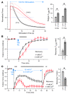
Figure 8. Mutant (+/m) EDL fatigued more slowly than control (+/+) muscle and was more vulnerable to impaired recovery in elevated [K+]o.
(AC) Fatigue was induced by continuous 100-Hz stimulation to isolated EDL muscles using 1-ms pulses in bath that contained 4 mM [K+] and 2 mM [Ca2+]. (A) In the left panel, tetanic force was normalized to the peak value, and the responses (mean ± SD, dashed lines) are shown for EDL from (+/m) mice (n = 8, 10.8 ± 2.2 months old) or (+/+) mice (n = 5, 11.4 ± 1.8 months old). The time required for decline to 50% of the peak force (fatigue T1/2R) was increased by 2.3-fold for the older mutant mice versus controls and by 1.8-fold for younger mutant mice (n = 6, 4.0 ± 0.7 months old) versus controls (n = 10, 4.2 ± 0.8 months old). (B) Recovery from fatigue for EDL muscles from 8.5 ± 0.4–month-old mutant (n = 6) and sibling controls (n = 5) in normal [K+]. Tetanic stimuli (0.5-ms, 70-mA current pulses for 300 ms at 125 Hz) were applied before and after 100-Hz stimulation (small blue box), and the normalized responses (mean ± SEM) are shown. The time required to regain 50% of the full extent of recovery (recovery T1/2R) was increased by 2.8-fold for mutant compared with control. (C) Stimulation of EDL muscles to fatigue as in B was followed by exposure to a bath containing 10 mM [K+] and 0.5 mM [Ca2+], which impaired the extent of recovery for mutant more than control muscles. Weakness was reversible in recovery buffer, and mutant muscles regained force 1.9-fold faster than did control muscle. Gray bars in the left panels indicate significant differences by ANOVA (P < 0.05) between mutant (red circles) and control (open squares) responses. The bar graphs in the right panels show mean ± SEM. *P < 0.005, 2-tailed Student’s t test.

Similar articles

Cited by

References

    1. Lehmann-Horn, F., Rudel, R., and Jurkat-Rott, K. 2004. Nondystrophic myotonias and periodic paralyses.Myology . A.G. Engel and C. Franzini-Armstrong, editors. 3rd edition. McGraw-Hill. New York, New York, USA. 1257–1300.
    1. Cannon S.C. Pathomechanisms in channelopathies of skeletal muscle and brain. Annu. Rev. Neurosci. 2006;29:387–415. doi: 10.1146/annurev.neuro.29.051605.112815. - DOI - PubMed
    1. Creutzfeldt O.D., Abbott B.C., Fowler W.M., Pearson C.M. Muscle membrane potentials in episodic adynamia. Electroencephalogr. Clin. Neurophysiol. 1963;15:508–519. doi: 10.1016/0013-4694(63)90071-3. - DOI - PubMed
    1. Tyler F.H., Stephens F.E., Gunn F.D., Perkoff G.T. Studies in disorders of muscle. VII. Clinical manifestations and inheritance of a type of periodic paralysis without hypopotassemia. . J. Clin. Invest. 1951;30:492–502. doi: 10.1172/JCI102465. - DOI - PMC - PubMed
    1. Gamstorp I., Hauge M., Helweglarsen H.F., Mjones H., Sagild U. Adynamia episodica hereditaria: a disease clinically resembling familial periodic paralysis but characterized by increasing serum potassium during the paralytic attacks. Am. J. Med. 1957;23:385–390. doi: 10.1016/0002-9343(57)90318-2. - DOI - PubMed

Publication types

MeSH terms