. 2008 Apr;47(4):1211-22.
doi: 10.1002/hep.22202.

Sulfatase 2 up-regulates glypican 3, promotes fibroblast growth factor signaling, and decreases survival in hepatocellular carcinoma

Affiliations

Sulfatase 2 up-regulates glypican 3, promotes fibroblast growth factor signaling, and decreases survival in hepatocellular carcinoma

Jin-Ping Lai et al. Hepatology. 2008 Apr.

Abstract

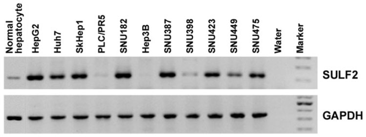
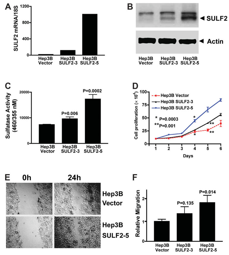
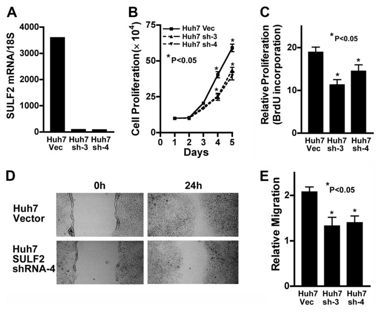
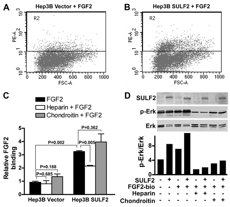
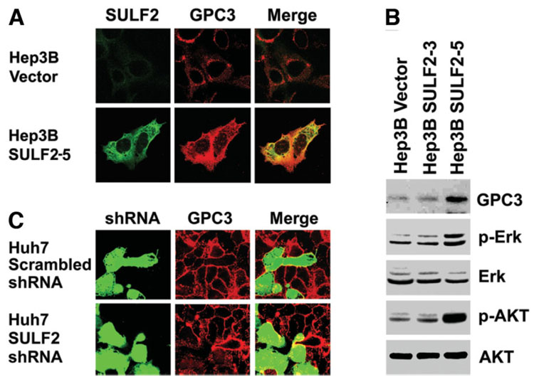
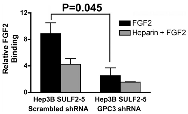
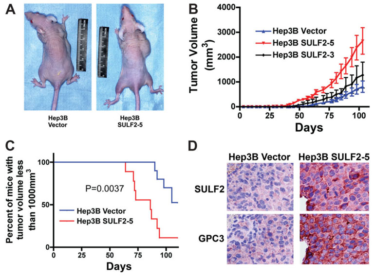
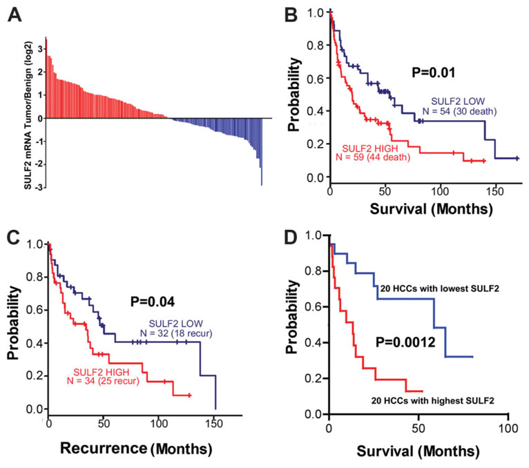
It has been shown that the heparin-degrading endosulfatase, sulfatase 1 (SULF1), functions as a liver tumor suppressor, but the role of the related sulfatase, sulfatase 2 (SULF2), in liver carcinogenesis remains to be elucidated. We investigated the effect of SULF2 on liver tumorigenesis. Expression of SULF2 was increased in 79 (57%) of 139 hepatocellular carcinomas (HCCs) and 8 (73%) of 11 HCC cell lines. Forced expression of SULF2 increased HCC cell growth and migration, whereas knockdown of SULF2 using short hairpin RNA targeting SULF2 abrogated HCC cell proliferation and migration in vitro. Because SULF1 and SULF2 desulfate heparan sulfate proteoglycans (HSPGs) and the HSPG glypican 3 (GPC3) is up-regulated in HCC, we investigated the effects of SULF2 on GPC3 expression and the association of SULF2 with GPC3. SULF2-mediated cell growth was associated with increased binding of fibroblast growth factor 2 (FGF2), phosphorylation of extracellular signal-regulated kinase and AKT, and expression of GPC3. Knockdown of GPC3 attenuated FGF2 binding in SULF2-expressing HCC cells. The effects of SULF2 on up-regulation of GPC3 and tumor growth were confirmed in nude mouse xenografts. Moreover, HCC patients with increased SULF2 expression in resected HCC tissues had a worse prognosis and a higher rate of recurrence after surgery.

Conclusion: In contrast to the tumor suppressor effect of SULF1, SULF2 has an oncogenic effect in HCC mediated in part through up-regulation of FGF signaling and GPC3 expression.


Conflict of interest statement

Potential conflict of interest: Nothing to report.

Figures

Fig. 1
Fig. 1. SULF2 mRNA expression in normal primary hepatocytes and 11 HCC cell lines measured by RT-PCR
GAPDH was used as a loading control.
Fig. 2
Fig. 2. Forced expression of SULF2 increases HCC cell proliferation and migration
(A) Plasmids expressing the full-length SULF2 mRNA or the control empty vector pcDNA3.1 were stably transfected into Hep3B cells. SULF2 mRNA expression in different clones was confirmed with quantitative real-time RT-PCR in the three indicated clones. Compared to Hep3B Vector cells, Hep3B SULF2-3 cells showed an intermediate level of SULF2 mRNA expression, whereas Hep3B SULF2-5 cells showed a high level of SULF2 mRNA expression. (B) Western immunoblotting was performed on whole cell lysates from Hep3B Vector, Hep3B SULF2-3, and Hep3B SULF2-5 cells with antibodies against SULF2; the blots were then stripped and reprobed with antibodies against actin to control for loading. (C) Nonsteroid sulfatase activity was detected in the indicated clones after inhibition of steroid sulfatases with estrone-3-O-sulfamate. (D) One hundred thousand Hep3B Vector, Hep3B SULF2-3, or Hep3B SULF2-5 cells were plated into 6-well plates, and 3 wells of each cell clone were harvested, stained with trypan blue, and counted each day. The mean and standard error of triplicate measurements are plotted (P < 0.01 at days 4, 5, and 6, Hep3B SULF2-5 versus Hep3B Vector). (E) Scratch wounds were made in confluent cell culture monolayers with a 20-µL pipette tip; photomicrographs of the wounds were taken at 0 and 24 hours thereafter. (F) Relative migration of the wound edge was quantitated and showed a significantly increased rate of migration in Hep3B SULF2-5 cells when compared to Hep3B Vector cells (P = 0.013).
Fig. 3
Fig. 3. Knockdown of SULF2 decreases HCC cell proliferation and migration
(A) Plasmid constructs expressing an shRNA targeting the SULF2 mRNA or the empty vector plasmid pSS-H1p were stably transfected into Huh7 cells, which expressed high levels of SULF2 mRNA. SULF2 mRNA levels were quantitated by real-time RT-PCR in Huh7 Vector (Huh7 Vec) and the two SULF2 shRNA clones, designated Huh7 SULF2shRNA-3 (Huh7 sh-3) and Huh7 SULF2shRNA-4 (Huh7 sh-4). SULF2 mRNA levels were profoundly suppressed by the shRNA plasmid. (B) Huh7 Vec, Huh7 sh-3, and Huh7 sh-4 cells were plated into 6-well plates at 1 × 10⁵ cells per well for the indicated time periods, and the total number of viable cells from each well was counted after trypan blue staining (P < 0.05 at days 4 and 5). (C) Huh7 Vec, Huh7 sh-3, and Huh7 sh-4 cells were plated into 96-well plates at 3000 cells per well. After 24 hours, cell proliferation was evaluated by the BrdU incorporation assay. Knockdown of SULF2 mRNA significantly decreased BrdU incorporation of both shRNA clones (P < 0.05). (D,E) Scratch wounds were made in confluent cell culture monolayers with a 20-µL pipette tip; photomicrographs of the wounds were taken at 0 and 24 hours thereafter. Relative migration of the wound edge was quantitated and showed a significantly decreased rate of migration in Huh7 sh-3 and Huh7 sh-4 cells when compared to Huh7 Vec cells (P < 0.05).
Fig. 4
Fig. 4. SULF2 increases FGF2 binding to the cell surface and activates the FGF signaling pathway
(A–C) Hep3B Vector and Hep3B SULF2-5 cells were collected and incubated with 10 nM biotinylated FGF2 ligand with or without 50 ng/mL heparin or 20 ng/mL chondroitin for 45 minutes at 4°C. After staining with streptavidin, 20,000 cells were counted by flow cytometry. (D) Hep3B Vector and Hep3B SULF2-5 cells were plated into 6-well plates and stimulated with 1 ng/mL biotinylated FGF2 for 15 minutes after overnight starvation. Western immunoblotting was performed on whole cell lysates with antibody against SULF2, p-Erk, and Erk. Optical densities of p-Erk and Erk were quantitated, and the ratios of p-Erk to Erk were calculated. Similar results were found after reprobing of the blots with p-AKT and AKT (data not shown).
Fig. 5
Fig. 5. Expression of SULF2 up-regulates cell-surface GPC3 and activates the Erk and AKT signaling pathways
(A) Hep3B Vector and Hep3B SULF2-5 cells were plated on cover slips in 6-well plates. Cells were stained with antibodies against SULF2 (green) and GPC3 (red) without permeabilization. SULF2 and GPC3 were both expressed at the cell surface and colocalized with each other. (B) The blots shown in Fig. 2B were stripped and reprobed with antibodies against GPC3, phospho-AKT, and phospho-Erk. Total AKT and total Erk were used as loading controls. (C) Huh7 cells were plated onto cover slips in 6-well plates and transiently transfected with GFP-expressing plasmids coexpressing either a scrambled shRNA sequence or an shRNA targeting SULF2 mRNA. After 24 hours, cells were immunostained for GPC3.
Fig. 6
Fig. 6. Knockdown of GPC3 inhibits FGF2 binding
Plasmid constructs expressing a GFP-coexpressing plasmid with an shRNA targeting the GPC3 mRNA or a scrambled shRNA were transiently transfected into Hep3B SULF2-5 cells. Cells were collected and incubated with 10 nM biotinylated FGF2 ligand with or without 50 ng/mL heparin for 45 minutes at 4°C. After staining with streptavidin, 20,000 cells were counted by flow cytometry. FGF2 binding was compared in GFP-gated cells.
Fig. 7
Fig. 7. SULF2 promotes tumor growth and up-regulates GPC3 in vivo in HCC xenografts in nude mice
(A) 5 × 10⁵ Hep3B Vector (left mouse) or Hep3B SULF2-5 cells (right mouse) in 150 µL of PBS were inoculated subcutaneously into the right frank of 10 mice for each cell clone. (B,C) Tumor growth was monitored with calipers. SULF2 expression significantly enhanced tumor growth in vivo. (D) Successive sections from paraffin-embedded xenografts from Hep3B Vector (left panel) and Hep3B SULF2-5 cells (right panel) were immunostained with antibodies against SULF2 and GPC3, respectively; nuclei were counterstained with hematoxylin.
Fig. 8
Fig. 8. SULF2 mRNA expression is increased in approximately 60% of HCCs
High SULF2 is associated with decreased patient survival and more rapid recurrence after surgical resection. (A) Results of oligonucleotide microarray analysis performed on 139 pairs of tumor and benign samples from primary HCCs, plotted as a log 2–fold change of SULF2 mRNA expression, tumor over benign. (B,C) Survival and tumor recurrence after surgical resection in 139 patients with HCCs. Differences between the Kaplan-Meier curves of patients with up-regulated expression of SULF2 and those with down-regulated expression of SULF2 with the log rank test. (D) Survival after surgical resection in the 20 patients with the highest tumor/benign SULF2 mRNA expression and in the 20 patients with the lowest tumor/benign SULF2 mRNA expression. Increased SULF2 is associated with a worse survival and more rapid recurrence after surgical resection of the primary tumor.

References

    1. Parkin DM, Bray F, Ferlay J, Pisani P. Global cancer statistics, 2002. CA Cancer J Clin. 2005;55:74–108. - PubMed
    1. El-Serag HB, Rudolph KL. Hepatocellular carcinoma: epidemiology and molecular carcinogenesis. Gastroenterology. 2007;132:2557–2576. - PubMed
    1. Roberts LR, Gores GJ. Emerging drugs for hepatocellular carcinoma. Expert Opin Emerg Drugs. 2006;11:469–487. - PubMed
    1. Abou-Alfa GK, Schwartz L, Ricci S, Amadori D, Santoro A, Figer A, et al. Phase II study of sorafenib in patients with advanced hepatocellular carcinoma. J Clin Oncol. 2006;24:4293–4300. - PubMed
    1. Morimoto-Tomita M, Uchimura K, Werb Z, Hemmerich S, Rosen SD. Cloning and characterization of two extracellular heparin-degrading endosulfatases in mice and humans. J Biol Chem. 2002;277:49175–49185. - PMC - PubMed

Publication types