. 2008 Mar 4;5(3):e54.
doi: 10.1371/journal.pmed.0050054.

Aberrant mucin assembly in mice causes endoplasmic reticulum stress and spontaneous inflammation resembling ulcerative colitis

Affiliations

Aberrant mucin assembly in mice causes endoplasmic reticulum stress and spontaneous inflammation resembling ulcerative colitis

Chad K Heazlewood et al. PLoS Med. .

Abstract

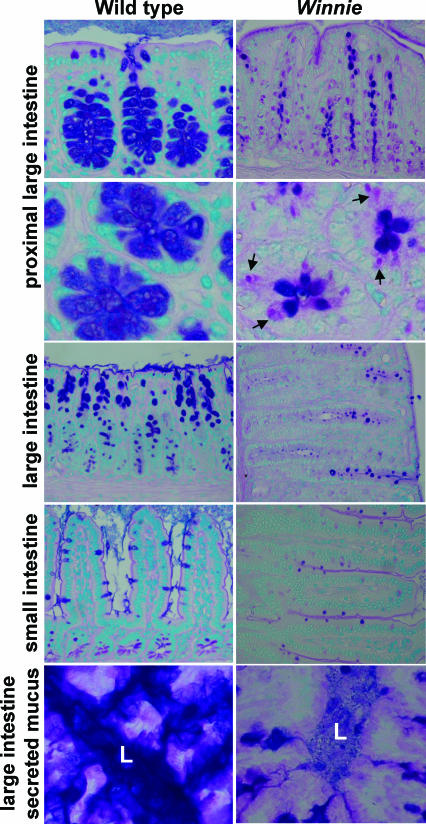
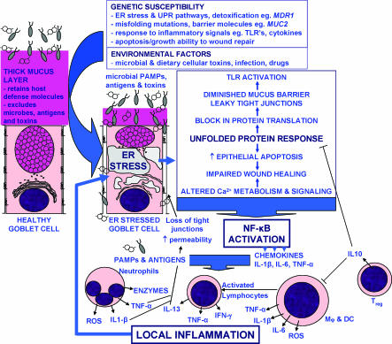
Background: MUC2 mucin produced by intestinal goblet cells is the major component of the intestinal mucus barrier. The inflammatory bowel disease ulcerative colitis is characterized by depleted goblet cells and a reduced mucus layer, but the aetiology remains obscure. In this study we used random mutagenesis to produce two murine models of inflammatory bowel disease, characterised the basis and nature of the inflammation in these mice, and compared the pathology with human ulcerative colitis.

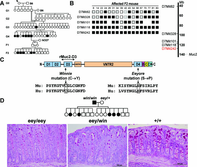
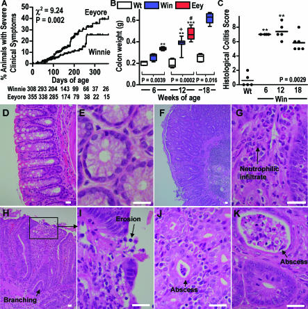
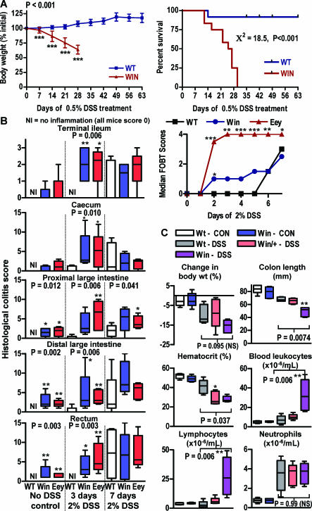
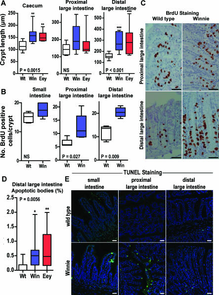
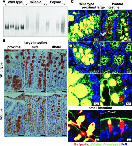
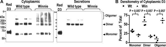
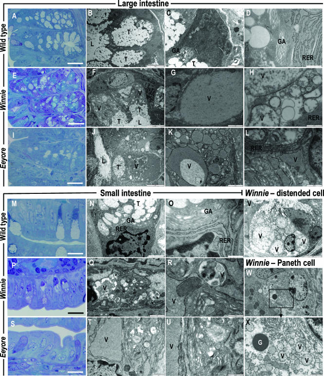
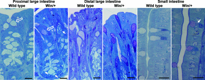
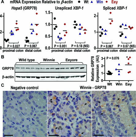
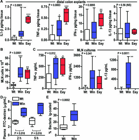
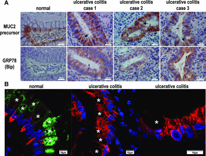
Methods and findings: By murine N-ethyl-N-nitrosourea mutagenesis we identified two distinct noncomplementing missense mutations in Muc2 causing an ulcerative colitis-like phenotype. 100% of mice of both strains developed mild spontaneous distal intestinal inflammation by 6 wk (histological colitis scores versus wild-type mice, p < 0.01) and chronic diarrhoea. Monitoring over 300 mice of each strain demonstrated that 25% and 40% of each strain, respectively, developed severe clinical signs of colitis by age 1 y. Mutant mice showed aberrant Muc2 biosynthesis, less stored mucin in goblet cells, a diminished mucus barrier, and increased susceptibility to colitis induced by a luminal toxin. Enhanced local production of IL-1beta, TNF-alpha, and IFN-gamma was seen in the distal colon, and intestinal permeability increased 2-fold. The number of leukocytes within mesenteric lymph nodes increased 5-fold and leukocytes cultured in vitro produced more Th1 and Th2 cytokines (IFN-gamma, TNF-alpha, and IL-13). This pathology was accompanied by accumulation of the Muc2 precursor and ultrastructural and biochemical evidence of endoplasmic reticulum (ER) stress in goblet cells, activation of the unfolded protein response, and altered intestinal expression of genes involved in ER stress, inflammation, apoptosis, and wound repair. Expression of mutated Muc2 oligomerisation domains in vitro demonstrated that aberrant Muc2 oligomerisation underlies the ER stress. In human ulcerative colitis we demonstrate similar accumulation of nonglycosylated MUC2 precursor in goblet cells together with ultrastructural and biochemical evidence of ER stress even in noninflamed intestinal tissue. Although our study demonstrates that mucin misfolding and ER stress initiate colitis in mice, it does not ascertain the genetic or environmental drivers of ER stress in human colitis.

Conclusions: Characterisation of the mouse models we created and comparison with human disease suggest that ER stress-related mucin depletion could be a fundamental component of the pathogenesis of human colitis and that clinical studies combining genetics, ER stress-related pathology and relevant environmental epidemiology are warranted.


Conflict of interest statement

Competing Interests: The authors have declared that no competing interests exist.

Figures

Figure 1
Figure 1. Histological Phenotype of Mice with Muc2 Mutations
PAS/Alcian blue stained intestinal sections from Winnie and wild-type C57BL/6 mice. Note the reduced size of Alcian blue staining thecae (stored mucin) and the presence of PAS-positive/Alcian blue negative accumulations (arrows) in Winnie goblet cells. L, lumen.
Figure 2
Figure 2. Generation and Characterization of Mice with Muc2 Mutations
(A) Dendrogram showing genesis of the Winnie mutation; filled symbols indicate the diarrhoea phenotype. (B) Microsatellite genotype on Chromosome 7 of 17 affected Winnie F2 mice. (C) Domain organization of the Muc2 protein showing the N- and C-terminal vWF D-domains (D1–D4), the C-terminal B, C, and CK domains, and the two central glycosylated tandem repeat domains (VNTR). The sites of the Winnie and Eeyore mutations are shown, together with the region cloned to express the rMuc2-D3 recombinant protein. (D) Results of complementation cross of Win/Win and Eey/+. PAS-stained sections of representative distal large intestinal histological phenotypes demonstrate noncomplementation (preservation of the eey/eey histological phenotype in eey/win).
Figure 3
Figure 3. Spontaneous Colitis in Mice with Muc2 Mutations
(A) Incidence of premature death or colitis-associated pathology requiring humane killing (chiefly rectal prolapse) in Winnie (n = 309) and Eeyore (n = 355) mice. Mice entering experiments or killed for other reasons were treated as censored observations (designated by upward ticks), and the number of uncensored mice remaining at 50 d intervals is included underneath the graph. Incidence rates in the two strains were compared using the Mantel Log-rank test. (B) Weight of the colon after removal of luminal faecal material in C57BL/6 (WT), Winnie (Win), and Eeyore (Eey) mice at 6 (Eey 6–9 wk), 12, and 18 (WT and Win only) wk of age, n = 4–9; box plots show median, quartiles, and range. (C) Histological colitis scores (see Materials and Methods) in WT and Win mice at 6, 12, and 18 wk of age, n = 4–6; scores from individual mice are shown. (D–K) Histology of normal distal colon from a C57BL/6 mouse (D and E) and examples of inflammation in the rectum (F–I) and distal large intestine (J and K) of untreated Winnie mice showing leukocytic infiltration (G and I), occasional branching crypts (F and H), crypt abscesses (J and K) and focal ulcerations (I); scale bars = 20 μm. Note the layer covering the mucosal surface in (F) is a granulocytic serous exudate. Statistics (B and C): p-values for Kruskal-Wallis nonparametric analysis are shown, Dunn's multiple comparison test versus wild type, ** p < 0.01, *** p < 0.001; versus Win at 6 wk, + p < 0.05; versus Eey at 6 wk, # p < 0.05.
Figure 4
Figure 4. Susceptibility of Mice with Muc2 Mutations to Dextran Sodium Sulphate–Induced Colitis
(A) Wild-type (WT) C57BL/6 and Winnie (Win) mice (n = 12) were given 0.5% DSS in drinking water for 63 d. Body weight (mean ± standard deviation, ANOVA p-value shown, Bonferroni's post-hoc test versus wild-type mice, *** p < 0.001) and overall survival (Kaplan-Meier survival estimates and Mantel log-rank test [χ2-value]) are shown. (B) Wild-type C57BL/6 (WT), and Winnie (Win), and Eeyore (Eey) mice not manifesting rectal bleeding or prolapse (n = 6) were given 2% DSS in drinking water or water alone for 7 d. Histological colitis scores were determined on days 3 and 7 and faecal occult blood determined daily. Statistics: box plots show median, quartiles, and range; p-values for Kruskal-Wallis nonparametric analysis are shown, Dunn's multiple comparison test versus WT, * p < 0.05, ** p < 0.01, *** p < 0.001. (C) Wild-type C57BL/6, Winnie, and Winnie heterozygous (Win/+) mice (n = 5) were given 3% DSS in drinking water or drinking water alone (CON) for 7 d, at which time body weight, colon length, and haematology were assessed. Statistics: box plots show median, quartiles, and range; p-values for Kruskal-Wallis nonparametric analysis comparing DSS-treated WT, Win/+, and Win mice are shown, Dunn's multiple comparison test versus WT, * p < 0.05, ** p < 0.01; NS, not significant. Additional data from these experiments are shown in Figures S2 and S3.
Figure 5
Figure 5. Intestinal Proliferation and Apoptosis Are Increased in Mice with Muc2 Mutations
(A) Length of caecal, proximal, and distal large intestinal crypts in wild-type C57BL/6 (WT, n = 12), Winnie (Win, n = 11), and Eeyore (Eey, n = 14) mice. (B) Assessment of incorporation of BrdU in the small intestine, and proximal and distal large intestine of WT (n = 5) and Win (n = 5) mice. For each region of the intestine in each mouse the number of BrdU-positive nuclei was counted in ten crypts (see Materials and Methods). (C) Representative examples of BrdU staining in the proximal and distal colon. (D) Increases in the percentage of apoptotic bodies in distal large intestinal crypts of the same mice as in (A). (E) TUNEL staining showing increased apoptosis in the small and large intestine of Winnie mice (images representative of three mice in each group). Scale bars (C and E) = 50 μm. Statistics: box plots show median, quartiles, and range; (A, B, and D) Kruskal-Wallis nonparametric analysis (p-values shown) with Dunn's multiple comparison test (*p < 0.05, ** p < 0.01, *** p < 0.001); (B) Mann-Whitney U-test, p-values shown. NS, not significant.
Figure 6
Figure 6. Altered Muc2 Protein Expression in Mice with Muc2 Mutations
(A) Muc2 proteins from wild-type, Winnie, and Eeyore intestinal extracts were reduced and separated by agarose gel electrophoresis and detected by Western blotting with the mM2.2 antibody. Note the reduced intensity and the altered electrophoretic migration in the mutant proteins. (B) Cellular localization of Muc2 in wild-type and Winnie colon tissue determined by immunohistochemistry with affinity purified mM2.2 antibody. Note the decreased size of goblet cell thecae and cytoplasmic Muc2 staining outside thecae in Winnie; scale bars = 25 μm. (C) Confocal microscopy of wild-type and Winnie goblet cells from the proximal colon and small intestine showing vacuolar accumulation of Muc2 (red) lacking O-linked sugars identified with the DBA lectin (green) in Winnie only. Goblet cell thecae appear yellow due to merging of the red Muc2 core peptide with the green O-linked sugar. Nuclei are stained with DAPI (blue). Scale bars as marked. Single colour images of these composites are provided in Figure S5A–S5C.
Figure 7
Figure 7. Altered Muc2 N-Terminal Oligomerisation Caused by the Win Mutation
(A) PAGE/Western blotting analysis of oligomerisation of the rMuc2-D3 wild-type proteins and proteins with the Win mutation in MKN45 cells and secretions following transfection (n = 4 separate transfections for each group). The position of the origin (o) and markers are shown. A reduced sample (D3 Red) on each gel shows the migration of the D3 monomer. Recombinant proteins were detected with the M2 anti-FLAG antibody, and no reactivity was seen with untransfected cells. Note the hyperoligomerisation of the Winnie D3 domain intracellularly and its failure to be secreted. (B) Densitometric analysis of oligomerisation of the rMuc2-D3 Winnie and wild-type proteins in MKN-45 cellular lysates following transfection. Relative expression of monomer, dimer, and higher-order oligomers was determined by densitometry and expressed as a percentage of the total densitometric value for each sample (lane). Statistics: individual data points and p-values from Mann-Whitney U-tests shown.
Figure 8
Figure 8. Ultrastructural Evidence of Endoplasmic Reticulum Stress in the Intestinal Epithelium of Mice with Muc2 Mutations
Semi-thin sections from resin embedded large intestine of wild-type (A), Winnie (E), and Eeyore (I) mice and small intestine of wild-type (M), Winnie (P), and Eeyore (S) mice stained with toluidine blue. Transmission electron micrographs from the large intestine (B–D, F–H, J–L, and V) and small intestine (N, O, Q, R, T, U, W, and X) of wild-type (B–D, N, and O), Winnie (F–H, Q, R, and V), and Eeyore (J–L, T, and U) mice. Note the reduced size of goblet cell thecae (indicated by a T) containing stored mucin granules and the presence of vacuoles (indicated by a V) surrounded by rough endoplasmic reticulum (RER) in Winnie and Eeyore. Other abbreviations: G, Paneth cell granule; GA, Golgi apparatus; L, lumen. Scale bars 40 μm (A, E, and I), 20 μm (L, O, and R), 5 μm (B, F, J, and U), 2 μm (C, G, K, M, P, S, and V), 1 μm (W), 50 nm (D, H, L, N, Q, and T).
Figure 9
Figure 9. Evidence of Mucin Misfolding in Heterozygous Mice
Intestinal tissue from mice heterozygous for the Win mutation (Win/+) was fixed with glutaraldehyde, embedded in resin, and semi-thin sections stained with toluidine blue. Note the variable degree of vacuolisation of the goblet cells in the base of the crypts in the proximal large intestine (solid white arrows) and the lack of/minimal vacuolisation in surface goblet cells in the proximal large intestine (hollow arrows), distal large intestine and small intestinal villi. Tissue from wild-type C57BL6 mice is shown for comparison. Scale bars = 20 μm.
Figure 10
Figure 10. Evidence of ER Stress and UPR Activation in Mice with Muc2 Mutations
(A) mRNA expression of hspa5 (GRP78) and the unspliced and spliced forms of Xbp-1 in the proximal and distal colon of wild-type C57BL/6 (WT), Winnie (Win), and Eeyore (Eey) mice determined by quantitative PCR and expressed relative to β-actin with the mean ratio for the WT group in each tissue corrected to 1. Statistics: n = 3 individual data points shown, Kruskal-Wallis nonparametric analysis (p-values shown) with Dunn's multiple comparison test (*p < 0.05 versus wild type). NS, not significant. (B) Fluorescence Western blotting was used to determine the expression of GRP78 in the proximal colons of WT, Win, and Eey mice. Blots were simultaneously stained with antibodies reactive with β-actin as a loading control, and densitometry was used to quantify expression of each protein, which was then expressed as a proportion of the mean expression in wild-type mice with the mean ratio of the WT group corrected to 1. Statistics as in (A), n = 5. (C) Transverse crypt section (centre) and a longitudinal crypt section through the vacuole-rich base of goblet cells (right) from Winnie mice showing immunohistochemical detection of GRP78 in vacuolar accumulations (solid arrows) but not in thecae where present (hollow arrows). Lack of staining in the absence of the GRP78 antibody demonstrates specificity (left, negative control). Scale bars = 50 μm.
Figure 11
Figure 11. Immunopathology in Mice with Muc2 Mutations
(A) Concentrations of IL-1β, TNF-α, IFN-γ, and IL-13 in supernatants from distal colonic explants cultured for 24 h from WT (n = 14), Win (n = 12), and Eey (n = 5) mice at 12–18 wk of age. (B) The number of leukocytes in the MLNs was determined in C57BL/6 (WT, n = 16), Winnie (Win, n = 10), and Eeyore (Eey, n = 5) mice at 12 wk of age. (C) Concentrations of TNF-α, IFN-γ, and IL-13 in supernatants from 2 × 10⁶ MLN leukocytes stimulated with PMA and ionomycin and cultured for 48 h, from WT (n = 11), Win (n = 9), and Eey (n = 5) mice at 12–18 wk of age. For cytokine analysis samples below the sensitivity of each assay were given values of 0. (D) Plasma FITC-dextran concentrations in WT and Win mice (n = 5) 2 and 5 h following oral gavage. (E) The proportion of bacteria coated with immunoglobulin determined by flow cytometry in faecal samples from WT (n = 10) and Win (n = 10) mice. Statistics: box plots show median, quartiles, and range; (A and C) Kruskal-Wallis nonparametric analysis (p-values shown) with Dunn's multiple comparison test (*p < 0.05, ** p < 0.01, ***p < 0.001 versus WT); NS, not significant; (B, D, and E) Mann-Whitney U-test, p-values shown.
Figure 12
Figure 12. Evidence for MUC2 Precursor Accumulation and ER Stress in Human UC
(A) The MUC2 precursor and GRP78 were detected immunohistochemically. One normal colonic biopsy and three representative cases of UC of ten examined are shown. Note the MUC2 precursor antibody does not react with goblet cell thecae, where present (arrows). (B) Confocal microscopy of goblet cells from the colon of an unaffected individual and two UC patients showing accumulation of the MUC2 precursor identified with antibody 4F1 (red) throughout the cytoplasm in UC goblet cells, many of which lack thecae, but not in normal colon. Note that 4F1 does not react with goblet cell thecae (identified by *) in normal or UC colon, demonstrating it cannot react with O-glycosylated MUC2 that has exited the Golgi apparatus. Note that some goblet cell thecae contain MUC2 with O-linked sugars identified with the DBA lectin (green) but that UC goblet cells are mostly DBA lectin-negative. Nuclei are stained with DAPI (blue). Single colour images of these composites are provided in Figure S7. Scale bars = 20 μm (A) and 10 μm (B).
Figure 13
Figure 13. A Model for ER Stress in the Pathophysiology of Intestinal Inflammation
This model recognizes that genetic and environmental factors can combine to invoke ER stress, reduced mucus production, and inflammation. Local inflammation can then exacerbate ER stress, potentially establishing a chronic cycle of inflammation in susceptible individuals. Abbreviations: ER, endoplasmic reticulum; Mϕ, macrophage; MDR1, multi-drug resistance gene 1; PAMP, prokaryocyte-associated molecular pattern; ROS, reactive oxygen species; TLR, toll-like receptor; UPR, unfolded protein response.

References

    1. Herrmann A, Davies JR, Lindell G, Martensson S, Packer NH, et al. Studies on the “insoluble” glycoprotein complex from human colon. Identification of reduction-insensitive MUC2 oligomers and C-terminal cleavage. J Biol Chem. 1999;274:15828–15836. - PubMed
    1. Lidell ME, Johansson ME, Morgelin M, Asker N, Gum JR, et al. The recombinant C-terminus of the human MUC2 mucin forms dimers in Chinese-hamster ovary cells and heterodimers with full-length MUC2 in LS 174T cells. Biochem J. 2003;372:335–345. - PMC - PubMed
    1. Godl K, Johansson ME, Lidell ME, Morgelin M, Karlsson H, et al. The N terminus of the MUC2 mucin forms trimers that are held together within a trypsin-resistant core fragment. J Biol Chem. 2002;277:47248–47256. - PubMed
    1. Asker N, Axelsson MAB, Olofsson SO, Hansson GC. Dimerization of the human MUC2 mucin in the endoplasmic reticulum is followed by a N-glycosylation-dependent transfer of the mono- and dimers to the Golgi apparatus. J Biol Chem. 1998;273:18857–18863. - PubMed
    1. Sheehan JK, Thornton DJ, Howard M, Carlstedt I, Corfield AP, et al. Biosynthesis of the MUC2 mucin—evidence for a slow assembly of fully glycosylated units. Biochem J. 1996;315:1055–1060. - PMC - PubMed

Publication types

MeSH terms