. 2008 Apr 25;283(17):11073-7.
doi: 10.1074/jbc.C700242200. Epub 2008 Mar 3.

Direct interaction between SET8 and proliferating cell nuclear antigen couples H4-K20 methylation with DNA replication

Affiliations

Direct interaction between SET8 and proliferating cell nuclear antigen couples H4-K20 methylation with DNA replication

Michael S Y Huen et al. J Biol Chem. .

Abstract

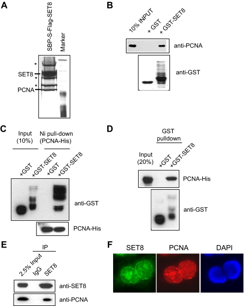
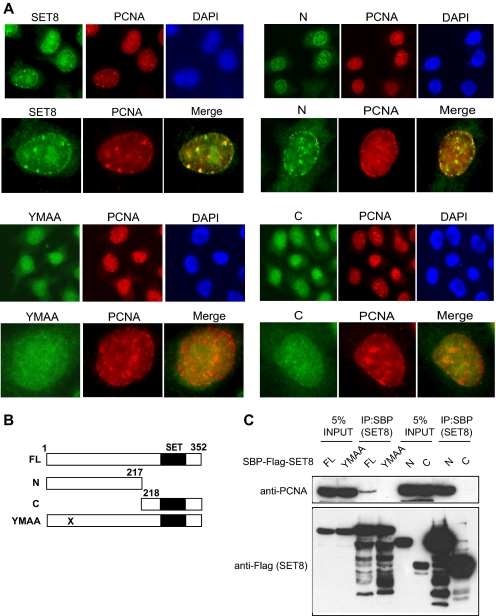
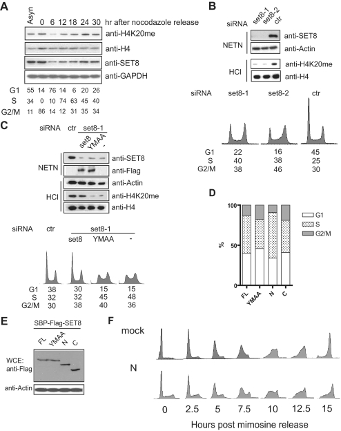
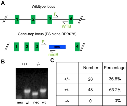
Chromatin endowed by histone modifications governs chromatin structure, which in turn represents a means to regulate cellular processes, including transcription and heterochromatin formation. Recent evidence revealed a plethora of enzymes that catalyze specific histone modifications for epigenetic maintenance, and dysregulation of which contributes to tumorigenesis and developmental defects. The histone methyltransferase SET8 (also known as Pr-Set7) was previously reported to monomethylate Lys(20) of histone H4. However, the temporal and spatial control of SET8 activity remains elusive. Here, we provide evidence to support that SET8 monomethylates Lys(20) of histone H4 during S phase by tethering to proliferating cell nuclear antigen via a putative proliferating cell nuclear antigen-interacting protein box. In addition, we show that SET8 function is required for S phase progression. Finally, deletion of SET8 in mice causes embryonic lethality, suggesting that SET8 plays an important role in mammalian embryogenesis.


Figures

FIGURE 1.
FIGURE 1.
SET8 interacts with PCNA in vitro and in vivo. A, identification of PCNA as a SET8-interacting partner. Shown is a silver stain of SET8-associated proteins separated by SDS-PAGE. B, GST-SET8 but not GST alone interacts with PCNA. C and D, SET8 directly interacts with PCNA. E, SET8 interacts with PCNA in vivo. F, SET8 localizes with PCNA in replication foci. IP, immunoprecipitation; DAPI, 4′,6-diamidino-2-phenylindole.
FIGURE 2.
FIGURE 2.
SET8 interacts with PCNA and localizes to replication foci via a putative PIP box. A, immunofluorescence studies of full-length SET8, its N- and C-terminal truncation mutants, and the PIP mutant (YMAA) and their colocalization with PCNA foci. B, schematic illustration of full-length (FL) SET8 and its mutants. C, PCNA coprecipitates with full-length SET8 and its N-terminal mutant, but not the YMAA and C-terminal mutants. DAPI, 4′,6-diamidino-2-phenylindole; IP, immunoprecipitation.
FIGURE 3.
FIGURE 3.
SET8 is involved in S phase progression. A, expression profile of SET8 and H4-K20 monomethylation status during cell cycle progression. Histone H4 and glyceraldehyde-3-phosphate dehydrogenase (GAPDH) were used as loading controls. Asyn, asynchronized. B, depletion of SET8 accumulates cells in S/G2 phase, which coincides with down-regulated H4-K20 monomethylation. C, function of SET8 in S/G2 progression requires its interaction with PCNA. Reintroduction of siRNA-resistant SET8 but not YMAA restored normal cell cycle progression. D, FACS analysis of HeLa cells transiently transfected with set8 or its mutants. E, expression of SET8 and its mutants in transiently transfected HeLa cells. F, expression of a dominant-negative SET8 mutant results in improper S phase progression. ctr, control; FL, full-length; WCE, whole cell extract.
FIGURE 4.
FIGURE 4.
SET8 is required for mouse development. A, schematic illustration of the wild-type (wt) set8 genomic locus and the gene-trapped locus. WTB, wild-type reverse primer; neoB, neo reverse primer. B, PCR analysis of F1 offspring. C, genotyping results obtained with offspring from the breeding of heterozygous set8 mice. EC, embryonic stem cell.

References

    1. Berger, S. L. (2007) Nature 447 407–412 - PubMed
    1. Mellor, J. (2006) Cell 126 22–24 - PubMed
    1. Schreiber, S. L., and Bernstein, B. E. (2002) Cell 111 771–778 - PubMed
    1. Botuyan, M. V., Lee, J., Ward, I. M., Kim, J. E., Thompson, J. R., Chen, J., and Mer, G. (2006) Cell 127 1361–1373 - PMC - PubMed
    1. Benetti, R., Gonzalo, S., Jaco, I., Schotta, G., Klatt, P., Jenuwein, T., and Blasco, M. A. (2007) J. Cell Biol. 178 925–936 - PMC - PubMed

Publication types