

Simultaneous in vivo positron emission tomography and magnetic resonance imaging
- PMID: 18319342
- PMCID: PMC2268792
- DOI: 10.1073/pnas.0711622105
Simultaneous in vivo positron emission tomography and magnetic resonance imaging
Abstract
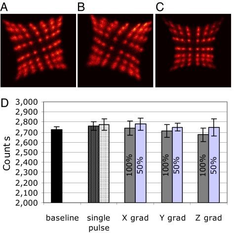
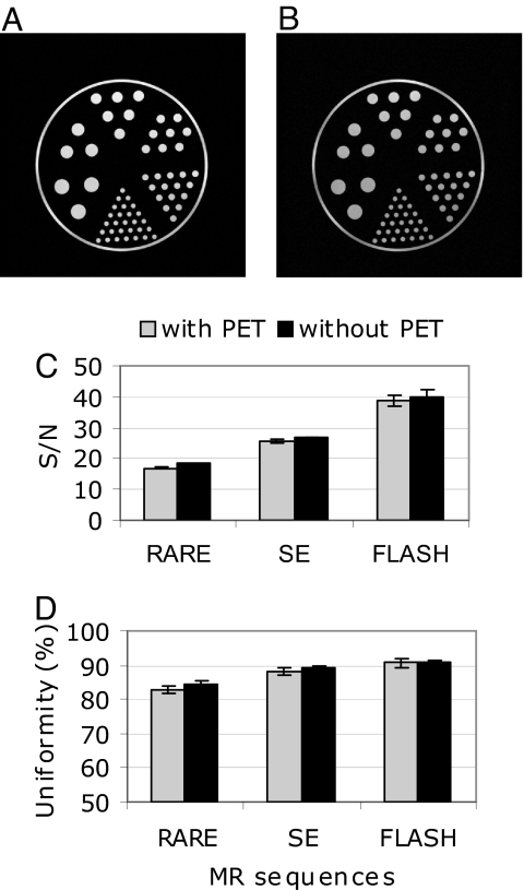
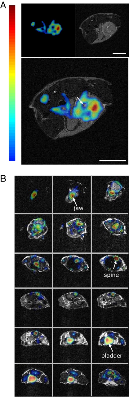
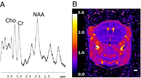
Positron emission tomography (PET) and magnetic resonance imaging (MRI) are widely used in vivo imaging technologies with both clinical and biomedical research applications. The strengths of MRI include high-resolution, high-contrast morphologic imaging of soft tissues; the ability to image physiologic parameters such as diffusion and changes in oxygenation level resulting from neuronal stimulation; and the measurement of metabolites using chemical shift imaging. PET images the distribution of biologically targeted radiotracers with high sensitivity, but images generally lack anatomic context and are of lower spatial resolution. Integration of these technologies permits the acquisition of temporally correlated data showing the distribution of PET radiotracers and MRI contrast agents or MR-detectable metabolites, with registration to the underlying anatomy. An MRI-compatible PET scanner has been built for biomedical research applications that allows data from both modalities to be acquired simultaneously. Experiments demonstrate no effect of the MRI system on the spatial resolution of the PET system and <10% reduction in the fraction of radioactive decay events detected by the PET scanner inside the MRI. The signal-to-noise ratio and uniformity of the MR images, with the exception of one particular pulse sequence, were little affected by the presence of the PET scanner. In vivo simultaneous PET and MRI studies were performed in mice. Proof-of-principle in vivo MR spectroscopy and functional MRI experiments were also demonstrated with the combined scanner.
Conflict of interest statement
The authors declare no conflict of interest.
Figures
References
-
- Phelps ME. PET: Molecular Imaging and Its Biological Applications. New York: Springer–Verlag; 2004. PET: Physics, instrumentation, and scanners. Chap 1.
-
- Gambhir SS. Molecular imaging of cancer with positron emission tomography. Nat Rev Cancer. 2002;2:683–693. - PubMed
-
- Gadian DG. NMR and Its Applications to Living Systems. New York: Oxford Univ Press; 1995.
-
- Toga AW, Mazziotta JC, editors. Brain Mapping: The Methods. 2nd Ed. San Diego: Academic; 2002.
Publication types
MeSH terms
Substances
Grants and funding
LinkOut - more resources
Full Text Sources
Other Literature Sources
Medical
