. 2008 Mar 5;3(3):e1708.
doi: 10.1371/journal.pone.0001708.

Signaling signatures and functional properties of anti-human CD28 superagonistic antibodies

Affiliations

Signaling signatures and functional properties of anti-human CD28 superagonistic antibodies

Zoe Waibler et al. PLoS One. .

Abstract

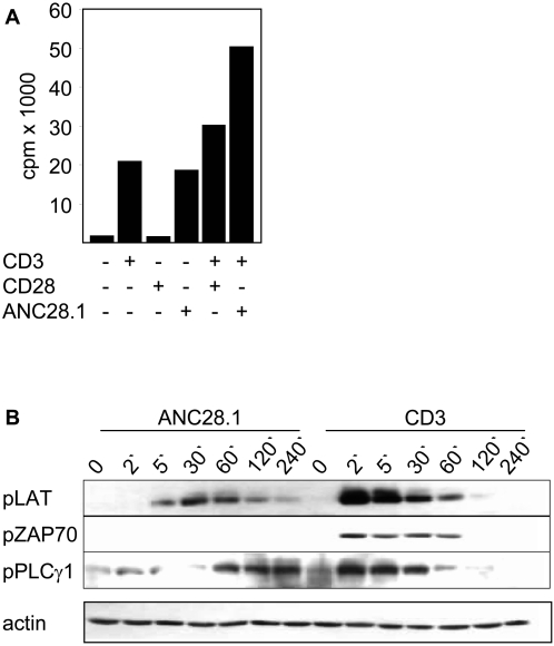
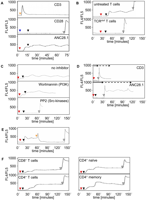
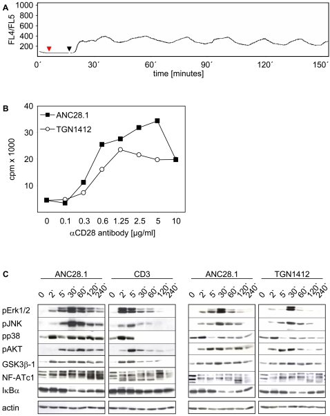
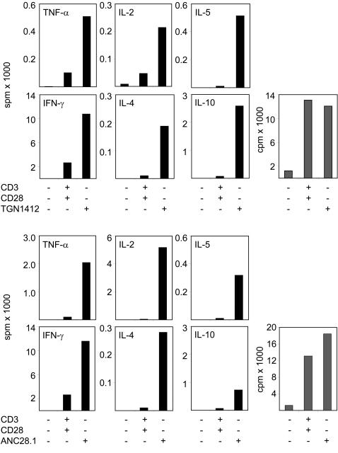
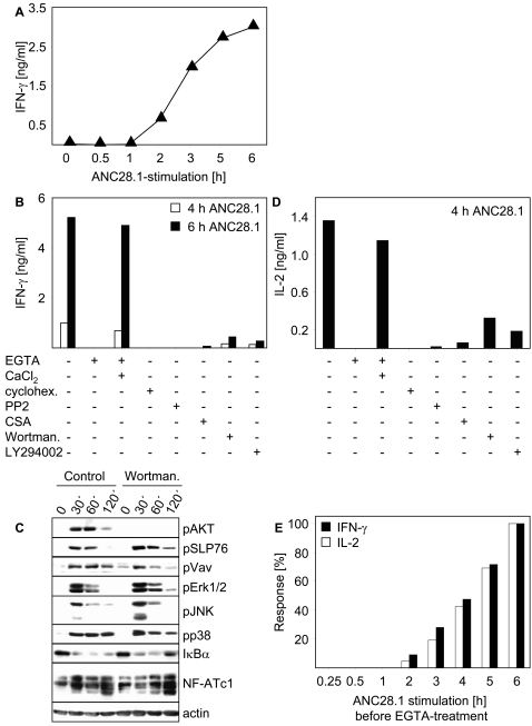
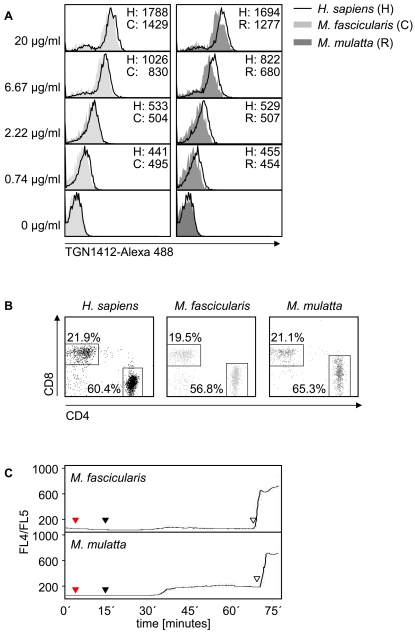
Superagonistic CD28 antibodies (CD28SAs) activate T lymphocytes without concomitant perturbation of the TCR/CD3-complex. In rodents these reagents induce the preferential expansion of regulatory T cells and can be used for the treatment of autoimmune diseases. Unexpectedly, the humanized CD28 superagonist TGN1412 caused severe and life threatening adverse effects during a recently conducted phase I clinical trail. The underlying molecular mechanisms are as yet unclear. We show that TGN1412 as well as the commercially available CD28 superagonist ANC28.1 induce a delayed but extremely sustained calcium response in human naïve and memory CD4+ T cells but not in cynomolgus T lymphocytes. The sustained Ca++-signal was associated with the activation of multiple intracellular signaling pathways and together these events culminated in the rapid de novo synthesis of high amounts of pro-inflammatory cytokines, most notably IFN-gamma and TNF-alpha. Importantly, sustained transmembranous calcium flux, activation of Src-kinases as well as activation of PI3K were found to be absolutely required for CD28SA-mediated production of IFN-gamma and IL-2. Collectively, our data suggest a molecular basis for the severe side effects caused by TGN1412 and impinge upon the relevance of non-human primates as preclinical models for reagents that are supposed to modify the function of human T cells.


Conflict of interest statement

Competing Interests: The authors have declared that no competing interests exist.

Figures

Figure 1
Figure 1. The CD28 superagonist ANC28.1 induces polyclonal T cell activation in vitro and leads to ZAP70 independent phosphorylation of LAT and PLCγ1.
(A) 5×10⁴ freshly prepared human T cells per well were seeded in 96-well plates and treated with the indicated combinations of antibodies or were incubated in medium as a control. 72 hours after stimulation, cultures were pulsed with 3H-thymidine for 6 hours and harvested thereafter. Note that ANC28.1 only induces polyclonal T cell proliferation when applied in soluble form (in this case 10 µg/ml of soluble ANC28.1 were crosslinked in solution with 20 µg/ml of polyclonal goat-anti-mouse antiserum). All other stimulations (including stimulation of T cells with CD3ε mAb and ANC28.1; very right bar in Fig. 1A) were performed in wells that were pre-coated with polyclonal goat anti-mouse antiserum. Shown data are representative for at least 6 independently performed experiments. (B) 1×10⁶ freshly prepared human T cells/lane were stimulated in solution with 10 µg/ml ANC28.1 followed by crosslinking with 20 µg/ml polyclonal goat anti-mouse antiserum for the indicated periods of time. As a control, cells were activated with a 1∶50 v/v dilution of ascites fluid of CD3ε mAb MEM92 (IgM) or were left untreated. Cell lysates were processed for western blotting using the indicated phosphospecific antibodies (Y319 of ZAP70, Y171 of LAT, and Y783 of PLCγ1). Western blotting using an anti-actin antibody was performed as loading control.
Figure 2
Figure 2. Induction of sustained calcium flux by human CD28 superagonistic antibody ANC28.1.
(A) 1×10⁶ freshly prepared Indo-1 loaded human T cells were stimulated with either a 1∶50 v/v dilution of ascites fluid of CD3 mAb MEM92 (orange triangle), 10 µg/ml conventional CD28 mAb (CD28.2; blue triangle) followed by crosslinking with 20 µg/ml polyclonal goat anti-mouse antiserum (black triangle) or 10 µg/ml ANC28.1 (red triangle) followed by crosslinking with 20 µg/ml polyclonal goat anti-mouse antiserum (black triangle). Induction of the Ca++-response was monitored for approximately 75 minutes. Note that using another conventional anti-CD28 mAb (248.23.2; with or without crosslinking) no Ca++-flux was induced as well (data not shown). (B) Untreated (untreated T cells) or 2AD2A2 pre-treated (TCRdull T cells) human T cells were stimulated with ANC28.1 (red triangles) and subsequently crosslinked (black triangles) as described in (A) and induction of the Ca++-response was monitored for the indicated periods of time. To confirm proper loading of the cells with Indo-1, the Ca++-ionophore Ionomycin (10 µg/ml) was added at the end of the experiment (unfilled triangle). (C) Cells were treated with ANC28.1 (red triangles) followed by crosslinking (black triangles) as described in (A) in the absence (no inhibitor) or presence of the PI3-kinase inhibitor Wortmannin (0.2 µM) or the Src-kinase inhibitor PP2 (10 µM). (D) CD3 (orange triangle) or ANC28.1 (red triangle) stimulation plus crosslinking (black triangle) was performed as described in (A) in culture medium that was supplemented with 1 mM EGTA to chelate extracellular Ca++ ions (solid arrow). At the indicated time points CaCl2 was added at a final concentration of 2 mM (dashed arrow). (E) Cells were treated with ANC28.1 as described in Fig. 1A. Additionally, at about 1 h of Ca-flux, a 1∶50 v/v dilution of ascites fluid of CD3 mAb MEM92 (orange triangle) was added. (F) CD8+, CD4+, CD4+/CD45RA+ naïve, and CD4+/CD45RO+ memory T lymphocytes were treated with ANC28.1 as described in (A) and induction of the Ca++-response was monitored for approximately 150 minutes.
Figure 3
Figure 3. Sustained calcium flux, T cell proliferation, and T cell signaling upon TGN1412 treatment.
(A) Freshly isolated human T cells were treated as described in Fig. 2 using the superagonistic CD28 specific mAb TGN1412 (10 µg/ml; red triangle). For crosslinking, 20 µg/ml of monoclonal mouse anti-human IgG4 was used (black triangle) and induction of the Ca++-response was monitored. (B) 5×10⁴ freshly prepared human T-cells/96-well were stimulated with the indicated concentrations of ANC28.1 or TGN1412 or were left untreated as a control. For crosslinking of ANC28.1, a polyclonal goat anti-mouse antiserum was used and crosslinking of TGN1412 was performed as described in (A). Concentrations for crosslinking were the following: 0.1–1.25 µg/ml primary Ab+2.5 µg/ml crosslinker; 2.5 µg/ml primary Ab+5 µg/ml crosslinker; 5 µg/ml primary Ab+10 µg/ml crosslinker; 10 µg/ml primary Ab+20 µg/ml crosslinker. 72 hours after stimulation, cultures were pulsed with 3H-thymidine for 6 hours, then harvested and proliferation analyzed by counts per minute [cpm]. (C) 1×10⁶ freshly prepared human T cells/lane were left untreated or were stimulated with 10 µg/ml ANC28.1 (first and third panel) followed by crosslinking with 20 µg/ml polyclonal goat anti-mouse antiserum or with 10 µg/ml TGN1412 (right panel) followed by crosslinking as described in (A) for the indicated periods of time. As a control, cells were activated with a 1∶50 dilution of ascites fluid of CD3 mAb MEM92 (second panel). Postnuclear lysates were processed for western blotting using the indicated phosphospecific antibodies. Western blotting using an anti-actin antibody was performed as loading control.
Figure 4
Figure 4. Cytokine production upon conventional and superagonistic CD28 stimulation of human T cells.
5×10⁴ freshly prepared human T-cells/96-well were stimulated with the indicated combination of antibodies as described in Fig. 1A or were left untreated as controls. At 48 hours 50 µl culture supernatant was removed from each well. Supernatants obtained from 3 wells were pooled and cytokine content was determined using the Cytokine Bead Array from BD in signals per minute [spm] (black bars). Additionally, induction of T cell proliferation was monitored upon treatment with the indicated antibody combinations (gray bars). 72 hours after stimulation, cultures were pulsed with 3H-thymidine for 6 hours, then harvested and proliferation analyzed by counts per minute [cpm]. Data represent the mean of 5 individual experiments.
Figure 5
Figure 5. Analysis of the signaling pathways controlling CD28SA-mediated IFN-γ and IL-2 production.
(A) Short term IFN-γ production upon CD28SA stimulation of T cells. 1×10⁶ T cells/200 µl were activated by CD28SA ANC28.1 as described for Figs. 1 –4. At the indicated time points 50 µl of the supernatants were removed, diluted 1∶3 v/v, and the concentration of secreted IFN-γ was determined using a commercially available ELISA. (B) Molecular requirements determining CD28SA-mediated IFN-γ production. T cells were pretreated for 30 minutes with the indicated substances prior to stimulation with ANC28.1. EGTA was used at 1 mM, CaCl2 at 2 mM, cycloheximide at 50 µg/ml, CSA at 150 ng/ml, PP2 at 10 µM, Wortmannin at 0.1 µM, and Ly294002 at 20 µM. At the indicated time points cytokine production was determined as described in (A). (C) 1×10⁶ freshly prepared human T cells/lane were stimulated with 10 µg/ml ANC28.1 followed by crosslinking with 20 µg/ml polyclonal goat anti-mouse antiserum with or without Wortmannin treatment. Western blotting was conducted as described in Fig. 3. Note that identical results were obtained when T cells were pretreated with LY294002 (data not shown). (D) Molecular requirements determining CD28SA-mediated IL-2 production. T cells were pretreated for 30 minutes with the indicated substances prior to stimulation with ANC28.1 as described in (B). 4 h after stimulation 50 µl of the supernatants were removed, diluted 1∶3 v/v, and the concentration of IL-2 was analyzed in supernatants by an ELISA. (E) CD28SA-mediated IFN-γ and IL-2 production requires sustained Ca++-flux. T cells were stimulated with ANC28.1 as described above. At 15′, 30′, 1 h, 2 h, 3 h, 4 h, and 5 h Ca++-flux was interrupted by addition of EGTA (1 mM) and the concentration of the cytokines in the supernatant was determined as described in (B) and (D). The concentrations of IL-2 and IFN-γ that were produced by ANC28.1-stimulated T cells following 6 hours of stimulation in the absence of EGTA were set to 100%. All data shown in Fig. 5 are representative for three independently performed experiments.
Figure 6
Figure 6. Despite similar TGN1412 binding, Macaca derived T cells show reduced calcium flux upon TGN1412 stimulation when compared to human T cells.
(A) MACS purified CD3+ human T cells (stained with an anti-CD3-APC antibody) and CD3+ monkey T cells (stained with an anti-CD3-PE antibody) were mixed 1∶1 and then incubated with graded concentrations of Alexa 488 labeled TGN1412. Binding of TGN1412-Alexa 488 was monitored by FACS analysis. Numbers given indicate the mean fluorescence intensity of samples. (B) Ratio of CD4+ vs. CD8+ T cells (gated on CD3+ cells) of human and monkey blood samples was analyzed by FACS analyses. (C) 1×10⁶ freshly isolated Indo-1 loaded M. fascicularis and M. mulatta T cells were treated as described in Fig. 2 using the superagonistic CD28 specific mAb TGN1412 (10 µg/ml; red triangle). For crosslinking, 20 µg/ml monoclonal mouse anti-human IgG4 was used (black triangle) and induction of the Ca++-response was monitored. To confirm viability of cells and proper loading with Indo-1, Ionomycin (10 µg/ml) was added at the end of the experiment (unfilled triangle).

References

    1. Schwartz RH. Costimulation of T lymphocytes: the role of CD28, CTLA-4, and B7/BB1 in interleukin-2 production and immunotherapy. Cell. 1992;71:1065–1068. - PubMed
    1. Sharpe AH, Abbas AK. T-cell costimulation–biology, therapeutic potential, and challenges. N Engl J Med. 2006;355:973–975. - PubMed
    1. Acuto O, Michel F. CD28-mediated co-stimulation: a quantitative support for TCR signalling. Nat Rev Immunol. 2003;3:939–951. - PubMed
    1. Rudd CE, Schneider H. Unifying concepts in CD28, ICOS and CTLA4 co-receptor signalling. Nat Rev Immunol. 2003;3:544–556. - PubMed
    1. Bischof A, Hara T, Lin CH, Beyers AD, Hunig T. Autonomous induction of proliferation, JNK and NF-alphaB activation in primary resting T cells by mobilized CD28. Eur J Immunol. 2000;30:876–882. - PubMed

Publication types

MeSH terms