

Early and late pathogenic events of newborn mice encephalitis experimentally induced by itacaiunas and curionópolis bracorhabdoviruses infection
- PMID: 18320052
- PMCID: PMC2253828
- DOI: 10.1371/journal.pone.0001733
Early and late pathogenic events of newborn mice encephalitis experimentally induced by itacaiunas and curionópolis bracorhabdoviruses infection
Abstract
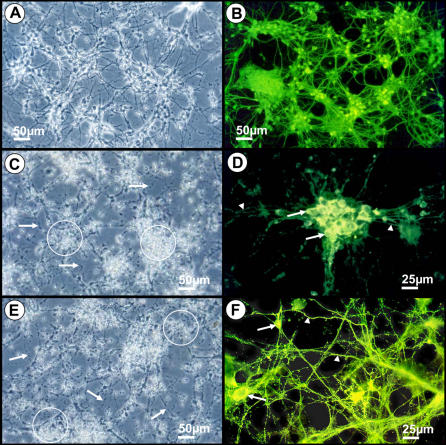
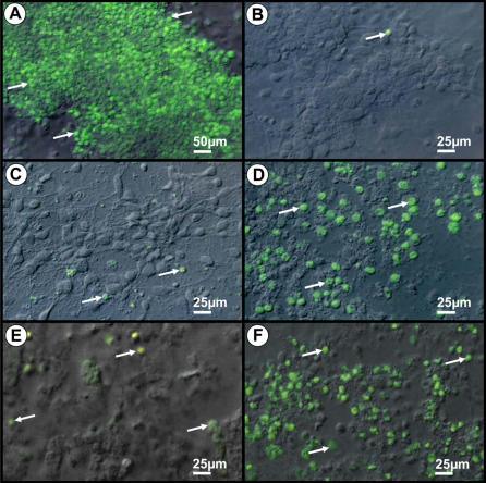
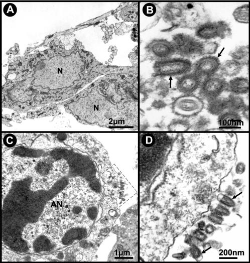
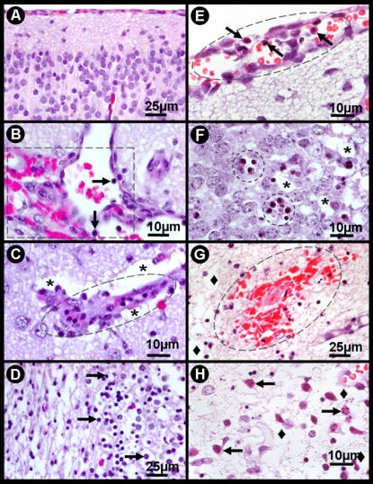
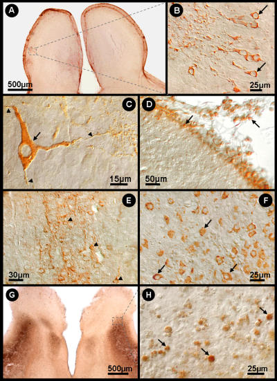
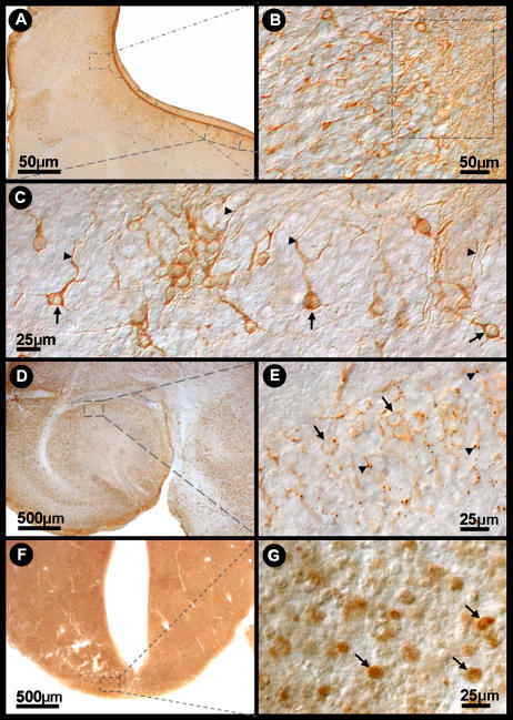
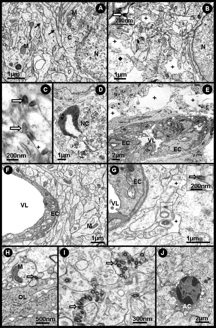
In previous reports we proposed a new genus for Rhabdoviridae and described neurotropic preference and gross neuropathology in newborn albino Swiss mice after Curionopolis and Itacaiunas infections. In the present report a time-course study of experimental encephalitis induced by Itacaiunas and Curionopolis virus was conducted both in vivo and in vitro to investigate cellular targets and the sequence of neuroinvasion. We also investigate, after intranasal inoculation, clinical signs, histopathology and apoptosis in correlation with viral immunolabeling at different time points. Curionopolis and Itacaiunas viral antigens were first detected in the parenchyma of olfactory pathways at 2 and 3 days post-inoculation (dpi) and the first clinical signs were observed at 4 and 8 dpi, respectively. After Curionopolis infection, the mortality rate was 100% between 5 and 6 dpi, and 35% between 8 and 15 dpi after Itacaiunas infection. We identified CNS mice cell types both in vivo and in vitro and the temporal sequence of neuroanatomical olfactory areas infected by Itacaiunas and Curionopolis virus. Distinct virulences were reflected in the neuropathological changes including TUNEL immunolabeling and cytopathic effects, more intense and precocious after intracerebral or in vitro inoculations of Curionopolis than after Itacaiunas virus. In vitro studies revealed neuronal but not astrocyte or microglial cytopathic effects at 2 dpi, with monolayer destruction occurring at 5 and 7 dpi with Curionopolis and Itacaiunas virus, respectively. Ultrastructural changes included virus budding associated with interstitial and perivascular edema, endothelial hypertrophy, a reduced and/or collapsed small vessel luminal area, thickening of the capillary basement membrane, and presence of phagocytosed apoptotic bodies. Glial cells with viral budding similar to oligodendrocytes were infected with Itacaiunas virus but not with Curionopolis virus. Thus, Curionopolis and Itacaiunas viruses share many pathological and clinical features present in other rhabdoviruses but distinct virulence and glial targets in newborn albino Swiss mice brain.
Conflict of interest statement
Figures
References
-
- Charles PC, Walters E, Margolis F, Johnston RE. Mechanism of neuroinvasion of Venezuelan equine encephalitis virus in the mouse. Virology. 1995;208:662–71. - PubMed
-
- Fazakerley JK. Semliki forest virus infection of laboratory mice: a model to study the pathogenesis of viral encephalitis. Arch Virol Suppl. 2004:179–90. - PubMed
-
- Jackson AC, Ye H, Ridaura-Sanz C, Lopez-Corella E. Quantitative study of the infection in brain neurons in human rabies. J Med Virol. 2001;65:614–8. - PubMed
-
- Saha S, Sugumar P, Bhandari P, Rangarajan PN. Identification of Japanese encephalitis virus-inducible genes in mouse brain and characterization of GARG39/IFIT2 as a microtubule-associated protein. J Gen Virol. 2006;87:3285–9. - PubMed
Publication types
MeSH terms
LinkOut - more resources
Full Text Sources
