. 2008 Mar;10(3):244-54.
doi: 10.1593/neo.07976.

CXCL5 promotes prostate cancer progression

Affiliations

CXCL5 promotes prostate cancer progression

Lesa A Begley et al. Neoplasia. 2008 Mar.

Abstract

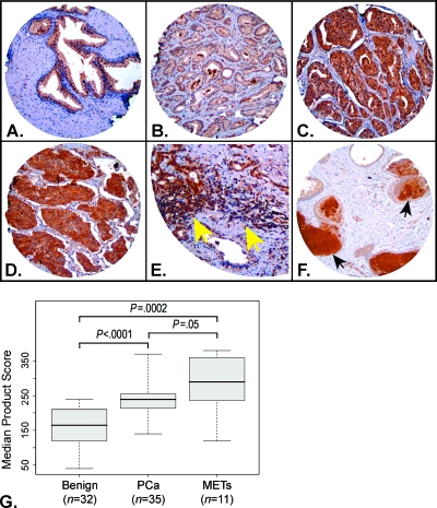
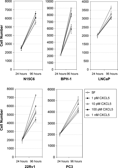
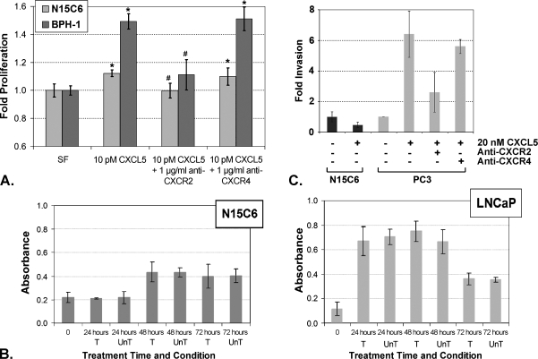
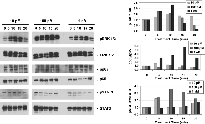
CXCL5 is a proangiogenic CXC-type chemokine that is an inflammatory mediator and a powerful attractant for granulocytic immune cells. Unlike many other chemokines, CXCL5 is secreted by both immune (neutrophil, monocyte, and macrophage) and nonimmune (epithelial, endothelial, and fibroblastic) cell types. The current study was intended to determine which of these cell types express CXCL5 in normal and malignant human prostatic tissues, whether expression levels correlated with malignancy and whether CXCL5 stimulated biologic effects consistent with a benign or malignant prostate epithelial phenotype. The results of these studies show that CXCL5 protein expression levels are concordant with prostate tumor progression, are highly associated with inflammatory infiltrate, and are frequently detected in the lumens of both benign and malignant prostate glands. Exogenous administration of CXCL5 stimulates cellular proliferation and gene transcription in both nontransformed and transformed prostate epithelial cells and induces highly aggressive prostate cancer cells to invade through synthetic basement membrane in vitro. These findings suggest that the inflammatory mediator, CXCL5, may play multiple roles in the etiology of both benign and malignant proliferative diseases in the prostate.


Figures

Figure 1
Figure 1
CXCL5 protein expression is concordant with prostate cancer progression. Shown are representative panels from a hematoxylin and eosin-stained, high-density tissue microarray probed with antibody against CXCL5, as follows: (A) Benign glands demonstrating weak staining. (B) PCa (Gleason sum 3 + 3) demonstrating weak staining. (C) PCa (Gleason sum 4 + 4) demonstrating moderate to strong staining. (D) Hormone refractory METs demonstrating strong staining. (E) PCa demonstrating moderate to strong staining associated with stromal inflammatory component (yellow arrows point to areas of inflammation). (F) Benign glands demonstrating strongly staining luminal secretions (black arrows). Original magnifications, x100. Panel E has been enlarged further, x4, to illustrate the area of inflammatory infiltrate concomitant with CXCL5 protein expression. (G) Boxplot depicting median product score distributions of protein expression levels for benign glands, malignant glands from PCa, and malignant areas from METs and P values associated with the statistical evaluation of these distributions.
Figure 2
Figure 2
Nontransformed and transformed prostate epithelial cells express the CXCL5 receptor and endogenously secrete CXCL5. (A) Immunoblot analysis of protein lysates prepared from transformed PC3 and LNCaP, and nontransformed N15C6 and BPH-1 prostate epithelial cells probed with antibodies specific for the CXCL5 receptor, CXCR2, and loading control, β-actin. Primary antibody concentrations used were 1:1000 for CXCR2 and 1:5000 for β-actin. (B) Protein levels (pg/ml) of CXCL5 present in media conditioned by transformed LNCaP and PC3 or nontransformed N15C6 or BPH-1 cells prostate epithelial cells were determined by ELISA. The graph shows the pg/ml CXCL5 detected plotted on a logarithmic scale (y axis).
Figure 3
Figure 3
Prostate epithelial cells proliferate in response to CXCL5. Nontransformed N15C6 or BPH-1 cells, androgen-sensitive transformed LNCaP or 22Rv1, or androgen-insensitive transformed PC3 cells, were plated at 1000 cells/well in triplicate in SF media, harvested, and counted 24 or 96 hours later. The mean and standard deviation (error bars) of cell numbers obtained at each time point for growth in SF media is indicated by white diamonds, in SF media supplemented to 1 pM CXCL5 by black diamonds, to 10 pM CXCL5 by white triangles, to 100 pM CXCL5 by black triangles, and to 1 nM CXCL5 by white squares. The experiments shown are representative of replicate proliferation assays.
Figure 4
Figure 4
CXCL5-stimulated proliferative and invasive responses. (A) N15C6 (light gray bars) or BPH-1 (dark gray bars) nontransformed prostate epithelial cells proliferated to significantly higher levels when grown for 72 hours in SF media supplemented with 10 pM CXCL5 than those grown in SF alone (*P < .001). Preincubation of the cells for 1 hour with 1 µg/ml antibody against CXCR2, the receptor for CXCL5, followed by supplementation with CXCL5 and maintenance of growth in CXCL5 + anti-CXCR2-containing media significantly ablated the proliferative response (#P < .001). In contrast, cellular growth after preincubation with an antibody against an unrelated chemokine receptor, CXCR4, followed by supplementation with CXCL5 and maintenance of growth in CXCL5 + anti-CXCR4-containing media was similar to that observed for non-pretreated cells grown in CXCL5-supplemented media and was significantly higher than that in SF alone (*P < .001). All data are shown normalized to growth in unsupplemented SF, which was set at one-fold. (B) N15C6 (LEFT) or LNCaP (RIGHT) cells were grown in SF media (untreated, UnT) or SF media supplemented with 10 pM CXCL5 for N15C6 or 100 pM CXCL5 for LNCaP (treated, T) for the times indicated. The cells were then harvested and assessed for nucleosomal DNA fragmentation. The fraction of cells exhibiting apoptosis plotted on the y axis was calculated as the difference in absorbance measured at 405 nm and at the reference wavelength of 490 nm after adjusting for background absorbance at both wavelengths. No significant differences in the fraction of cells exhibiting apoptosis were observed between treated and untreated cells at any time point, demonstrating that CXCL5 does not promote antiapoptotic responses in these cells. (C) Fifteen thousand each of N15C6 (black bars) or PC3 (gray bars) cells were plated onto Matrigel-coated membranes and were exposed to complete media or complete media supplemented with 20 nM CXCL5 for 24 hours. After 24 hours, the cells that migrated and invaded through the Matrigel were stained and counted. N15C6 cells did not demonstrate an invasive response to treatment with CXCL5. However, approximately six-fold more PC3 cells migrated through the synthetic basement membrane, Matrigel, in response to 20 nM CXCL5 compared to vehicle (control, set at one-fold) (*P < .05). PC3 cell invasion through the Matrigel in response to CXCL5 was significantly inhibited by pretreatment with 1 µg/ml blocking antibody (anti-CXCR2) (#P < .05) but not by pretreatment with nonspecific antibody (anti-CXCR4) (*P < .05).
Figure 5
Figure 5
CXCL5 activates MAPK signaling in nontransformed N15C6 prostate epithelial cells. Nontransformed N15C6 cells rapidly and transiently phosphorylated ERK 1/2 and STAT3 when treated with either subnanomolar (10 or 100 pM) or nanomolar (1 nM) levels of CXCL5, whereas NF-κB subunit activation was evident only after treatment with 1 nM CXCL5. Primary antibody concentrations used were 1:500 for phospho-ERK, 1:500 for phospho-65 (NF-κB), 1:1000 for phospho-STAT3, 1:1000 for total ERK, 1:1000 for total p65, and 1:2000 for total STAT3. A total of 20 µg of protein lysate was electrophoresed per well. Immunoblots are shown on the left, and corresponding densitometric evaluations of the same blots are shown on the right. Phosphorylation relative to total protein quantitated from the immunoblot is shown in the densitometric plots as phospho/total protein.
Figure 6
Figure 6
CXCL5 activates both MAPK and PI3K signaling in transformed LNCaP prostate epithelial cells. Transformed LNCaP cells rapidly and transiently phosphorylated both ERK 1/2 and the p65 subunit of NF-κB on treatment with subnanomolar (10 or 100 pM) levels of CXCL5. Immunoblots are shown in the top panel, and corresponding densitometric evaluations of the same blots are shown in the bottom panel. Phosphorylation relative to total protein quantitated from the immunoblot is shown in the densitometric plots as phospho/total protein. A total of 100 µg of protein lysate was electrophoresed per well. Primary antibody concentrations used were as described for Figure 5.
Figure 7
Figure 7
CXCL5 stimulates a transcriptional response in both nontransformed and transformed prostate epithelial cells. Quantitative real-time PCR of RNA purified from N15C6 cells (left) or LNCaP cells (right) treated with subnanomolar CXCL5 as shown demonstrates rapid and robust transcription of the EGR1 gene significantly higher than levels obtained at time 0 (set at one-fold) (*P < .05). Data shown are averaged from three or more separate experiments per time point per concentration of CXCL5 examined.

References

    1. Jemal A, Siegel R, Ward E, Murray T, Xu J, Thun MJ. Cancer statistics, 2007. CA Cancer J Clin. 2007;57:43–66. - PubMed
    1. Koch AE. Chemokines and their receptors in rheumatoid arthritis: future targets? Arthritis Rheum. 2005;52:710–721. - PubMed
    1. Begley L, Monteleon C, Shah RB, Macdonald JW, Macoska JA. CXCL12 overexpression and secretion by aging fibroblasts enhance human prostate epithelial proliferation in vitro. Aging Cell. 2005;4(6):291–298. - PubMed
    1. Begley LA, MacDonald JW, Day ML, Macoska JA. CXCL12 activates a robust transcriptional response in human prostate epithelial cells. J Biol Chem. 2007;282(37):26767–26774. - PubMed
    1. Singh S, Singh UP, Grizzle WE, Lillard JW., Jr CXCL12-CXCR4 interactions modulate prostate cancer cell migration, metalloproteinase expression and invasion. Lab Invest. 2004;84(12):1666–1676. [PMID: 15467730] - PubMed

Publication types

MeSH terms

Substances

LinkOut - more resources