Review
. 2008 Apr;129(4):407-20.
doi: 10.1007/s00418-008-0406-y. Epub 2008 Mar 5.

Autophagy-physiology and pathophysiology

Affiliations
Review

Autophagy-physiology and pathophysiology

Yasuo Uchiyama et al. Histochem Cell Biol. 2008 Apr.

Abstract

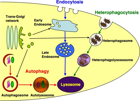
"Autophagy" is a highly conserved pathway for degradation, by which wasted intracellular macromolecules are delivered to lysosomes, where they are degraded into biologically active monomers such as amino acids that are subsequently re-used to maintain cellular metabolic turnover and homeostasis. Recent genetic studies have shown that mice lacking an autophagy-related gene (Atg5 or Atg7) cannot survive longer than 12 h after birth because of nutrient shortage. Moreover, tissue-specific impairment of autophagy in central nervous system tissue causes massive loss of neurons, resulting in neurodegeneration, while impaired autophagy in liver tissue causes accumulation of wasted organelles, leading to hepatomegaly. Although autophagy generally prevents cell death, our recent study using conditional Atg7-deficient mice in CNS tissue has demonstrated the presence of autophagic neuron death in the hippocampus after neonatal hypoxic/ischemic brain injury. Thus, recent genetic studies have shown that autophagy is involved in various cellular functions. In this review, we introduce physiological and pathophysiological roles of autophagy.


Figures

Fig. 1
Fig. 1
Biogenesis of lysosomes: cells execute receptor-mediated endocytosis, heterophagocytosis, and autophagy, forming early endosomes, heterophagosomes, and autophagosomes, respectively. To degrade ingested materials, each structure receives lysosomal enzymes by fusing with transporting vesicles from TGN or lysosomes and becomes late endosomes, heterophagolysosomes, and auto(phago)lysosomes. Before receiving lysosomal enzymes, autophagosomes fuse with endosomes and become amphisomes (Gordon and Seglen 1988) that are not drawn in this diagram
Fig. 2
Fig. 2
Diagram showing an autophagic pathway. Excess, old, and unneeded macromolecules, including long-lived proteins and organelles happen to be sequestered and enwrapped completely by the ER-like isolation membrane, the origin of which is unknown, and become autophagosomes that receive lysosomal enzymes by fusing with transporting vesicles from trans-Golgi network or lysosomes. Then degradation starts and autophagosomes become auto(phago)lysosomes. Lysosomes contain acid hydrolases such as cathepsins
Fig. 3
Fig. 3
Electron micrographs of hepatocytes obtained from a mouse housed under starvation conditions for 24 h. Numerous vacuolar structures (arrowheads) are detectable near bile canaliculi (a). Some of these vacuoles are enwrapped by double membranes with morphologically intact cytoplasm (b), and double- or single-membranes with degraded, but morphologically identifiable cytoplasmic materials and structures (ce). Some vacuoles are encircled by double membranes and a part or whole portion of the intermembrane space is occupied with a dense material (d, e). A lysosome has heterogeneously electron dense materials (f). Bars indicate 1 mm in a and 0.5 μm in bf
Fig. 4
Fig. 4
Electron micrographs of hepatocytes obtained from a mouse housed under starvation conditions for 48 h. Numerous vacuolar structures (arrowheads) are detectable near bile canaliculi. Vacuolar structures are clearly larger in hepatocytes from mice starved for 48 h (a) than from mice starved for 24 h (see Fig. 3a). Some of these vacuoles are enwrapped by double membranes with morphologically intact cytoplasm (b, c), and single membranes with degraded but morphologically identifiable cytoplasmic materials and structures (df). Bars indicate 1 μm in a and 0.5 μm in bf
Fig. 5
Fig. 5
Western blotting for LC3. PC12 cells were incubated in the absence of serum and in the presence of a cysteine proteinase inhibitor, E-64-d, or an aspartic proteinase inhibitor, pepstatin A, for 3, 6 or 12 h (h). Lysates from E-64-d-treated, pepstatin A-treated, and control untreated (before serum-free culturing) PC12 cells at each time point were subjected to western blotting. Protein bands immunoreactive for LC3 are detected at molecular weights of 18 and 16 kDa, which correspond to membrane-bound LC3-II and cytosolic LC3-I, respectively. The LC3-II form increases with time after the start of serum-free culturing, indicative of progression of the autophagic process
Fig. 6
Fig. 6
Immunostaining for LC3 in PC12 cells. Cells were cultured in the absence of serum and in the presence of E-64-d, a cysteine proteinase inhibitor, and/or pepstatin A, an aspartic proteinase inhibitor, for 3 or 6 h (h). Cells that were cultured in the presence of serum and in the absence of serum without inhibitors were used as control. Punctate signals for LC3 are distinct in cells treated with inhibitors. Bar indicates 10 μm
Fig. 7
Fig. 7
Immunoelectron microscopy for LC3 using the SDS-FRL method. Immunogold particles indicating LC3 are specifically present on the granule membranes. Bar indicates 0.5 μm
Fig. 8
Fig. 8
Immunostaining for ubiquitin (Ubi) in the liver of control (Atg7flox/flox) (a) and Atg7flox/flox; Mx1-Cre (b) mice. Mice were injected with pIpg once a week and killed 16 days after the first injection. Positive staining for ubiquitin is intensely detected in coarse granules in Atg7-deficient hepatocytes but not in the control ones. Bar indicates 20 μm
Fig. 9
Fig. 9
Immunostaining for ubiquitin (Ubi) in the cerebral cortex of control (Atg7flox/flox) (a) and Atg7flox/flox; Nestin-Cre (b) mice at 4 weeks of age. Positive signals for ubiquitin are intensely detected in cortical neurons of an Atg7-deficient brain, but not in the control cortical neurons. Bar indicates 10 μm
Fig. 10
Fig. 10
Immunostaining for LC3 in cathepsin D-deficient (CD−/−) (b) and littermate control (CD+/−) (a) mouse cerebral cortexes at P23. Positive signals for LC3 are intensely detected in granules of cortical neurons deficient in CD, while they are fibrilar in dendrites of both control and mutant neurons. Bar indicates 20 μm
Fig. 11
Fig. 11
Electron micrographs of cerebral cortical neurons in mouse brains deficient in cathepsin D (CD−/−) at 3,523 and doubly deficient in cathepsins B and L (CB−/−CL−/−) at P13. Granular osmiophilic deposits (GRODs) abundantly accumulate in neuronal perikarya of both mutant mouse brains. GRODs in the neurons are frequently enwrapped together with part of the cytoplasm by double-membrane structures. Bars indicate 1 μm

References

    1. {'text': '', 'ref_index': 1, 'ids': [{'type': 'DOI', 'value': '10.2353/ajpath.2006.051066', 'is_inner': False, 'url': 'https://doi.org/10.2353/ajpath.2006.051066'}, {'type': 'PMC', 'value': 'PMC1780162', 'is_inner': False, 'url': 'https://pmc.ncbi.nlm.nih.gov/articles/PMC1780162/'}, {'type': 'PubMed', 'value': '16877357', 'is_inner': True, 'url': 'https://pubmed.ncbi.nlm.nih.gov/16877357/'}]}
    2. Adhami F, Liao G, Morozov YM, Schloemer A, Schmithorst VJ, Lorenz JN, Dunn RS, Vorhees CV, Wills-Karp M, Degen JL, Davis RJ, Mizushima N, Rakic P, Dardzinski BJ, Holland SK, Sharp FR, Kuan CY (2006) Cerebral ischemia-hypoxia induces intravascular coagulation and autophagy. Am J Pathol 169:566–583 - PMC - PubMed
    1. {'text': '', 'ref_index': 1, 'ids': [{'type': 'PubMed', 'value': '17035724', 'is_inner': True, 'url': 'https://pubmed.ncbi.nlm.nih.gov/17035724/'}]}
    2. Adhami F, Schloemer A, Kuan CY (2007) The roles of autophagy in cerebral ischemia. Autophagy 3:42–44 - PubMed
    1. {'text': '', 'ref_index': 1, 'ids': [{'type': 'DOI', 'value': '10.1016/j.febslet.2004.12.058', 'is_inner': False, 'url': 'https://doi.org/10.1016/j.febslet.2004.12.058'}, {'type': 'PubMed', 'value': '15670810', 'is_inner': True, 'url': 'https://pubmed.ncbi.nlm.nih.gov/15670810/'}]}
    2. Ardley HC, Hung CC, Robinson PA (2005) The aggravating role of the ubiquitin–proteasome system in neurodegeneration. FEBS Lett 579:571–576 - PubMed
    1. {'text': '', 'ref_index': 1, 'ids': [{'type': 'PMC', 'value': 'PMC2013521', 'is_inner': False, 'url': 'https://pmc.ncbi.nlm.nih.gov/articles/PMC2013521/'}, {'type': 'PubMed', 'value': '4300890', 'is_inner': True, 'url': 'https://pubmed.ncbi.nlm.nih.gov/4300890/'}]}
    2. Arstila AU, Trump BF (1968) Studies on cellular autophagocytosis. The formation of autophagic vacuoles in the liver after glucagon administration. Am J Pathol 53:687–733 - PMC - PubMed
    1. {'text': '', 'ref_index': 1, 'ids': [{'type': 'DOI', 'value': '10.1083/jcb.12.1.198', 'is_inner': False, 'url': 'https://doi.org/10.1083/jcb.12.1.198'}, {'type': 'PMC', 'value': 'PMC2106008', 'is_inner': False, 'url': 'https://pmc.ncbi.nlm.nih.gov/articles/PMC2106008/'}, {'type': 'PubMed', 'value': '13862833', 'is_inner': True, 'url': 'https://pubmed.ncbi.nlm.nih.gov/13862833/'}]}
    2. Ashford TP, Porter KR (1962) Cytoplasmic components in hepatic cell lysosomes. J Cell Biol 12:198–202 - PMC - PubMed

Publication types

MeSH terms

LinkOut - more resources