. 2008 May;46(5):485-97.
doi: 10.1007/s11517-008-0315-1.

Alterations in vasomotor control of coronary resistance vessels in remodelled myocardium of swine with a recent myocardial infarction

Affiliations

Alterations in vasomotor control of coronary resistance vessels in remodelled myocardium of swine with a recent myocardial infarction

Dirk J Duncker et al. Med Biol Eng Comput. 2008 May.

Abstract

The mechanism underlying the progressive deterioration of left ventricular (LV) dysfunction after myocardial infarction (MI) towards overt heart failure remains incompletely understood, but may involve impairments in coronary blood flow regulation within remodelled myocardium leading to intermittent myocardial ischemia. Blood flow to the remodelled myocardium is hampered as the coronary vasculature does not grow commensurate with the increase in LV mass and because extravascular compression of the coronary vasculature is increased. In addition to these factors, an increase in coronary vasomotor tone, secondary to neurohumoral activation and endothelial dysfunction, could also contribute to the impaired myocardial oxygen supply. Consequently, we explored, in a series of studies, the alterations in regulation of coronary resistance vessel tone in remodelled myocardium of swine with a 2 to 3-week-old MI. These studies indicate that myocardial oxygen balance is perturbed in remodelled myocardium, thereby forcing the myocardium to increase its oxygen extraction. These perturbations do not appear to be the result of blunted beta-adrenergic or endothelial NO-mediated coronary vasodilator influences, and are opposed by an increased vasodilator influence through opening of K(ATP) channels. Unexpectedly, we observed that despite increased circulating levels of noradrenaline, angiotensin II and endothelin-1, alpha-adrenergic tone remained negligible, while the coronary vasoconstrictor influences of endogenous endothelin and angiotensin II were virtually abolished. We conclude that, early after MI, perturbations in myocardial oxygen balance are observed in remodelled myocardium. However, adaptive alterations in coronary resistance vessel control, consisting of increased vasodilator influences in conjunction with blunted vasoconstrictor influences, act to minimize the impairments of myocardial oxygen balance.


Figures

Fig. 1
Fig. 1
Alterations in determinants of oxygen supply and demand in remodelled myocardium in swine with a 3-week-old myocardial infarction (MI). The net effect of these alterations is a decrease in oxygen supply/demand ratio
Fig. 2
Fig. 2
Cardiovascular responses to exercise in normal swine and swine with a ∼3-week-old MI. 0L = lying, 0S = standing. Data are mean ± SEM; *P < 0.05 versus 0L, †P < 0.05 MI versus normal a corresponding exercise level. Data are from Haitsma et al. [43]
Fig. 3
Fig. 3
Neurohumoral responses in exercising swine with a ∼3-week-old MI. In the norepinephrine, epinephrine and dopamine panels, data shown for N represent 0L, 1, 2, 3, 4 and 5 km/h and data for MI represent 0L, 1, 2, 3 and 4 km/h. In all other panels, data shown for N represent 0L, 1, 3 and 5 km/h and data for MI represent 0L, 1, 3 and 4 km/h. Mean data points were fitted with second order curves. VO2 = O2-consumption. Data are mean ± SEM; *< 0.05 vs 0L, P < 0.05 MI versus Normal. Data are from Haitsma et al [43]
Fig. 4
Fig. 4
Myocardial blood flow and O2-balance in the left ventricular anterior wall of N and MI ∼3 weeks after myocardial infarction. Epi subepicardial, OM outer mid; IM inner mid; Endo subendocardial. MVO2 = myocardial O2-consumption; MEO2 = myocardial O2 extraction; CVPO2 = coronary venous O2 tension In the top left panel data myocardial blood flow data are shown for resting (Rest, lying) conditions, and during maximum exercise (Ex, 5 km/h in N and 4 km/h in MI). Data are mean ± SEM; *< 0.05 versus 0L, P < 0.05 MI versus Normal. Data are from Haitsma et al [43]
Fig. 5
Fig. 5
Effect of saline, α-adrenoceptor blockade (phentolamine, 1 mg/kg iv) and/or β-adrenoceptor blockade (propranolol, 0.5 mg/kg iv) and of muscarinic receptor blockade (atropine, 30 μg/kg/min, iv) and/or β-adrenoceptor blockade (propranolol, 0.5 mg/kg iv) on the response of coronary venous O2 tension (CVPO2) plotted as a function of myocardial O2 consumption (MVO2) in seven normal and 7 MI swine. *< 0.05 versus corresponding Control; < 0.05 Atropine + Propranolol versus corresponding Atropine or Phentolamine + Propranolol versus corresponding Phentolamine; Data are mean ± SEM. Data are from Duncker et al. [23]
Fig. 6
Fig. 6
Effect of AT1-receptor blockade with irbesartan (1 mg/kg iv) on the relation between myocardial O2 consumption (MVO2) and coronary venous O2 tension (CVPO2) in normal swine and swine with a 2–3-week-old myocardial infarction. Data are means ± SE; *< 0.05 versus corresponding control relation, < 0.05 effect of irbesartan different in MI versus Normal animals. Data are from Merkus et al. [68]
Fig. 7
Fig. 7
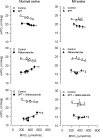
Effect of the adenosine receptor antagonist 8-phenyltheophylline (8PT, 5 mg/kg iv), the KATP channel blocker glibenclamide (Glib, 3 mg/kg iv) or their combination on myocardial O2 balance in the LV anterior free wall of normal swine and swine with a recent MI. MVO2 = myocardial O2 consumption; CVPO2 = coronary venous O2 tension; Data are mean ± SEM; Data are mean ± SEM; *P ≤ 0.05 versus corresponding control; P ≤ 0.05 effect of Glib was blunted at higher MVO2 levels (Glib × MVO2); P ≤ 0.05 effect of Glib different after MI (Glib × MVO2 × MI). Data are from Merkus et al. [71]
Fig. 8
Fig. 8
Effect of inhibition of NO synthase by NLA (20 mg/kg iv) on myocardial O2 extraction and coronary venous PO2 at rest (lying) and during treadmill exercise in MI and N. Data are mean ± SEM. *< 0.05 NLA versus corresponding Control; there were no significant differences in the responses to NLA between MI and N either at rest or during exercise. Data are from Haitsma et al. [44]
Fig. 9
Fig. 9
Effect of ETA receptor blockade with EMD (3 mg/kg iv) on myocardial oxygen balance in normal swine and swine with a recent MI. Data are means ± SE; *< 0.05 versus corresponding Control relation, < 0.05 effect of EMD waned during exercise. Data are from Merkus et al. [70]
Fig. 10
Fig. 10
Myocardial oxygen balance in normal and MI swine. Shown are the actual relations between MVO2 and CVPO2 in 30 normal swine (open circles) and 20 MI swine (open triangles) under control conditions. In addition, we have depicted the computed relations in MI swine if the ET (solid diamonds) and ANG II (solid squares) vasoconstrictor influences (which were both attenuated in MI swine) and the KATP (solid triangles) vasodilator influences (which were enhanced in MI swine) would have been identical to those in normal swine. The graph clearly illustrates that the adaptations in coronary vasomotor control act to blunt perturbations in oxygen balance in remodelled myocardium of swine with a recent MI
Fig. 11
Fig. 11
Alterations in vasomotor balance in the coronary resistance vessels within remodelled myocardium in swine with a 2–3-week-old myocardial infarction

Similar articles

Cited by

References

    1. Azevedo ER, Parker JD. Parasympathetic control of cardiac sympathetic activity: Normal ventricular function versus congestive heart failure. Circulation. 1999;100:274–279. doi: 10.1161/01.CIR.100.3.274. - DOI - PubMed
    1. Bache RJ. Effects of hypertrophy on the coronary circulation. Prog Cardiovasc Dis. 1988;30:403–440. doi: 10.1016/0033-0620(88)90005-9. - DOI - PubMed
    1. Bache RJ, Dai XZ, Schwartz JS, et al. Role of adenosine in coronary vasodilation during exercise. Circ Res. 1988;62:846–853. doi: 10.1161/01.RES.62.4.846. - DOI - PubMed
    1. Batenburg WW, Garrelds IM, Bernasconi CC, et al. Angiotensin II type 2 receptor-mediated vasodilation in human coronary microarteries. Circulation. 2004;109:2296–2301. doi: 10.1161/01.CIR.0000128696.12245.57. - DOI - PubMed
    1. Berne RM, Rubio R. Regulation of coronary blood flow. Adv Cardiol. 1974;12:303–317. doi: 10.1159/000395474. - DOI - PubMed

Publication types