. 2008 May 9;283(19):12797-810.
doi: 10.1074/jbc.M801122200. Epub 2008 Mar 5.

Cue1p is an activator of Ubc7p E2 activity in vitro and in vivo

Affiliations

Cue1p is an activator of Ubc7p E2 activity in vitro and in vivo

Omar A Bazirgan et al. J Biol Chem. .

Abstract

Ubc7p is a ubiquitin-conjugating enzyme (E2) that functions with endoplasmic reticulum (ER)-resident ubiquitin ligases (E3s) to promote endoplasmic reticulum-associated degradation (ERAD). Ubc7p only functions in ERAD if bound to the ER surface by Cue1p, a membrane-anchored ER protein. The role of Cue1p was thought to involve passive concentration of Ubc7p at the surface of the ER. However, our biochemical studies of Ubc7p suggested that Cue1p may, in addition, stimulate Ubc7p E2 activity. We have tested this idea and found it to be true both in vitro and in vivo. Ubc7p bound to the soluble domain of Cue1p showed strongly enhanced in vitro ubiquitination activity, both in the presence and absence of E3. Cue1p also enhanced Ubc7p function in vivo, and this activation was separable from the established ER-anchoring role of Cue1p. Finally, we tested in vivo activation of Ubc7p by Cue1p in an assay independent of the ER membrane and ERAD. A chimeric E2 linking Ubc7p to the Cdc34p/Ubc3p localization domain complemented the cdc34-2 TS phenotype, and co-expression of the soluble Cue1p domain enhanced complementation by this chimeric Ubc7p E2. These studies reveal a previously unobserved stimulation of Ubc7p E2 activity by Cue1p that is critical for full ERAD and that functions independently of the well known Cue1p anchoring function. Moreover, it suggests a previously unappreciated mode for regulation of E2s by Cue1p-like interacting partners.


Figures
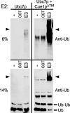
FIGURE 1.
FIGURE 1.
Cue1pΔ™ enhanced production of large and small ubiquitin-immunoreactive bands by Ubc7p. In vitro ubiquitination reactions with no E3 (–), GST (GST), or GST-Hrd1p (GST-E3) were prepared with either Ubc7p (left) or Ubc7p·Cue1pΔ™ (right). Identical samples were resolved by SDS-PAGE using 8% gels (top) or 14% gels (bottom), revealing the production of both large and small ubiquitin-immunoreactive bands. Ubiquitination was E3-dependent with Ubc7p alone, whereas Ubc7p·Cue1pΔ™ enhanced ubiquitination both in the presence and absence of E3 (compare left and right panels). The arrowheads indicate the discontinuity between 4% stacking gels and running gels. The molecular weights of monoubiquitin (Ub) and diubiquitin (Ub-Ub) are indicated.
FIGURE 2.
FIGURE 2.
Coomassie-stained in vitro ubiquitination reactions and E2s. Ubiquitination reactions without E3 and with Ubc7p, Ubc7p·Cue1pΔ™, HUBC4, or no E2, were resolved with 14% SDS-PAGE and Coomassie-stained to observe the size and quantity of small protein products produced by the in vitro ubiquitination reactions (Reactions). Purified E2 preparations were loaded on the same gel at 5 times higher concentration than in the ubiquitination reactions (E2 only [5×]) to identify those bands contributed by the E2 preparations. The molecular weights of monoubiquitin (Ub) and diubiquitin (Ub-Ub) are indicated. *, higher molecular weight bands produced in the Ubc7p·Cue1pΔ™ reaction.
FIGURE 3.
FIGURE 3.
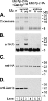
E3-indpendent ubiquitination produced multimers of ubiquitin linked through lysine 48. In vitro reactions with either Ubc7p-2HA· Cue1pΔ™ or Ubc7p-2HA were prepared with one of three versions of ubiquitin: wild-type ubiquitin (WT), ubiquitin with lysine 48 changed to arginine (K48R), or ubiquitin with all lysines except lysine 48 changed to arginine (K48only). Reactions were run and then split into four identical portions, resolved by 14% SDS-PAGE and Coomassie-stained or immunoblotted as indicated. E3-independent reaction products enhanced by Cue1p required lysine 48 of ubiquitin, and these higher molecular weight products were composed only of ubiquitin protein (anti-Ub), and not isopeptide conjugates to Ubc7p (anti-HA) or Cue1p (anti-Cue1p) in these reducing conditions.
FIGURE 4.
FIGURE 4.
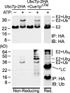
Polyubiquitin conjugates were thioester-linked to Ubc7p in vitro. In vitro reactions with either Ubc7p-2HA or Ubc7p-2HA·Cue1pΔ™ were immunoprecipitated with antibody-conjugated resin to HA epitope. Bound proteins were resolved by nonreducing SDS-PAGE (left) or reducing SDS-PAGE (right), and immunoblotted for HA epitope (top) or ubiquitin (bottom). Ubc7p-2HA showed an ATP-dependent shift in molecular weight in the nonreducing conditions. This shift was improved in the presence of Cue1pΔ™ and was reversed in reducing conditions. Ubiquitin immunoblotting showed high molecular weight ubiquitin chains co-precipitating in these nonreducing conditions. The ubiquitin dimer prevalent in Figs. 1, 2, 3 was not released from the E2 in nonreducing conditions but was liberated in the reducing conditions. The reducing conditions also released an antibody light chain (*LC) from the antibody-conjugated resin that was detected in the ubiquitin immunoblot.
FIGURE 5.
FIGURE 5.
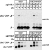
Cue1p did not strongly enhance the ubiquitin charging of Ubc7p by E1. In vitro assays of ubiquitin-Ubc7p thioester formation were performed with the indicated concentrations of Ubc7p-2HA (left lanes) or Ubc7p-2HA·Cue1pΔ™ (right lanes) and resolved with nonreducing SDS-PAGE. Ubiquitin immunoblotting (top) and HA epitope immunoblotting (bottom) both revealed a band the size of ubiquitin-Ubc7p thioester (Ubc7p-2HA-Ub) that diminished with E2 concentration. Ubc7p-2HA and Ubc7p-2HA·Cue1pΔ™ produced ubiquitin-Ubc7p thioester with similar efficiency at the lowest, most physiological E2 concentrations tested.
FIGURE 6.
FIGURE 6.
Biochemical analysis of Ubc7p and Ubc7p-2HA·Cue1pΔ™ in solution. A, Cue1p did not drastically modify the overall structure of Ubc7p. Limited trypsin proteolysis for the indicated times was performed on Ubc7p-2HA or Ubc7p-2HA·Cue1pΔ™, and then proteolyzed proteins were resolved by SDS-PAGE, and HA epitope immunoblotting revealed proteolysis of Ubc7p-2HA. The presence of Cue1p did not grossly modify the structure of Ubc7p in solution. The presence of the interacting Cue1pΔ™ provided Ubc7p-2HA some protection from proteolysis. However, this did not alter the molecular weights of the discrete bands that formed, suggesting that Ubc7p structure was intact in the absence of Cue1p. B, Cue1p and Ubc7p formed a dimer in solution, not a multimer of dimers. Superose-6 gel filtration chromatography of Ubc7p-2HA·Cue1pΔ™ was performed, and fractions were analyzed by SDS-PAGE. Coomassie staining revealed the peak elution fractions for the Ubc7p-2HA and Cue1pΔ™ proteins. Because these proteins were similar in size (∼20 kDa), we immunoblotted fraction samples to confirm the presence of both proteins. Peak fractions for protein-sizing standards, albumin (67 kDa), ovalbumin (43 kDa), chymotrypsin (25 kDa), and RNase A (13.7 kDa) were determined in identical conditions and are indicated with bars above the fraction numbers.
FIGURE 7.
FIGURE 7.
Partial restoration of ERAD in ubc7Δ strains by ER-anchored Ubc7p. Hmg2p-GFP levels were measured in ubc7Δ strains expressing either empty vector or the indicated ERAD proteins. Histograms plot the number of cells (y axis) having a given arbitrary fluorescence (x axis). A, Hmg2p is degraded in a wild-type strain (WT) and stabilized in a ubc7Δ null strain (ubc7Δ). B, Cue1p overexpression had no effect on ERAD. The histogram of a ubc7Δ null strain expressing Cue1p-3HA from the strong TDH3 promoter is superimposed on wild type and ubc7Δ histograms to test complementation of ubc7Δ. C, the histogram of a ubc7Δ null strain expressing membrane-anchored Ubc7p-2HA (TM-Ubc7p) reveals partial complementation of ubc7Δ.
FIGURE 8.
FIGURE 8.
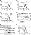
Soluble Cue1p domain enhanced ERAD by self-anchored TM-Ubc7p in vivo. Hmg2p-GFP levels were measured as above, in cue1Δ ubc7Δ strains with either empty vector or the indicated ERAD proteins. A, Cue1pΔ™ caused no reduction in Hmg2p-GFP levels. B, TM-Ubc7p caused little if any reduction in Hmg2p-GFP. C, when expressed together, TM-Ubc7p and Cue1pΔ™ allowed a reduction in Hmg2p-GFP levels, partially complementing the cue1Δ ubc7Δ double null. D, cycloheximide chase degradation assays were performed to confirm Hmg2p-GFP degradation in wild-type or cue1Δ ubc7Δ strains expressing either empty vector or the indicated ERAD proteins. Hmg2p-GFP levels were measured as above, and reduction of GFP fluorescence was plotted for each strain treated with 50μg/ml cycloheximide for 30 or 120 min. This confirmed that degradation of Hmg2p-GFP was enhanced only in the presence of both TM-Ubc7p and Cue1pΔ™. E, whole cell lysates and microsome fractions of the strains used in Fig. 8, A–D, reveal that TM-Ubc7p is localized to the microsomal membrane fraction, that co-expression of Cue1pΔ™ does not alter TM-Ubc7p levels, and that TM-Ubc7p recruits Cue1pΔ™ to the membrane fraction. F, L-Ubc7p, a version of Ubc7p with an N-terminal flexible linker, weakly complements Ubc7p function in comparison with Ubc7p.
FIGURE 9.
FIGURE 9.
A strategy to test Ubc7p function, independent of ERAD. A, primary sequence alignment of Ubc7p and Cdc34p. *, the conserved cysteine residue (Cys-89 in Ubc7p, Cys-95 in Cdc34p) where the ubiquitin thioester forms. Cdc34p has an acidic C-terminal tail required for localization to the SCF ubiquitin ligase complex. B, schematic of Ubc7p, Cdc34p, and a construct fusing the E2 domain of Ubc7p with the C-terminal tail of Cdc34p (Ubc7p-Cdc34) to target Ubc7p to the SCF ubiquitin ligase complex.
FIGURE 10.
FIGURE 10.
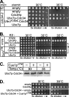
Cue1p enhanced Ubc7p activity in an ERAD-independent cellular context. A, restoration of Cdc34p function was assayed as complementation of the cdc34-2 temperature-sensitive growth phenotype. Wild-type strains (WT) grew at both 30 °C and 35 °C, but cdc34-2 allele strains do not survive at 35 °C. Cdc34p, Ubc7p, Ubc7p-Cdc34, and Ubc7pC89S-Cdc34 proteins were each expressed from the strong TDH3 promoter in the cdc34-2 strain. Expression of both Cdc34p and Ubc7p-Cdc34 restored the wild-type phenotype. This complementation was abolished when the conserved catalytic cysteine in Ubc7p-Cdc34 was mutated to serine (Ubc7pC89S-Cdc34). Fusion of Ubc7p to the Cdc34p tail domain was required for complementation of the cdc34-2 phenotype, as expression of Ubc7p alone had no effect. B, Cdc34p, Ubc7p, and Ubc7p-Cdc34 were expressed from the weaker CDC34 promoter in cdc34-2 allele strains. These strains also contained either empty vector (top four rows) or a vector expressing Cue1pΔ™ (bottom four rows). These strains were grown at permissive (30 °C) or nonpermissive (33 and 35 °C) temperatures. Here, Ubc7p-Cdc34 only slightly improved complementation of the TS phenotype compared with empty vector and was less effective than Cdc34p at complementing the cdc34-2 TS phenotype. Impressively, the addition of Cue1pΔ™ enhanced complementation only in the presence of Ubc7p-Cdc34, and not Cdc34p or Ubc7p. An asterisk highlights improved complementation by Ubc7p-Cdc34 upon the addition of Cue1pΔ™. C, levels of Ubc7p-Cdc34 were not changed by the addition of Cue1pΔ™. Whole cell lysates of strains grown for 5 h at either permissive (30 °C) or nonpermissive (33 °C) temperature were resolved by SDS-PAGE and immunoblotted for Ubc7p to examine Ubc7p-Cdc34 levels. At both permissive and nonpermissive temperatures, expression of Cue1pΔ™ caused no change in Ubc7p-Cdc34 levels. Thus, enhanced complementation by Cue1pΔ™ with Ubc7p-Cdc34 in Fig. 10B was due to increased activity of Ubc7p-Cdc34. D, using a cdc34-2 strain that was also cue1Δ, we observed complementation of the cdc34-2 TS phenotype by expression of Cdc34p from the CDC34 promoter (top). In this strain, we also observed partial TS rescue by expression of Ubc7p-Cdc34 from the CDC34 promoter, and enhancement of this complementation when Cue1pΔ™ is co-expressed. The similar trends in Fig. 10, B and D, with CUE1 or cue1Δ cdc34-2 strains suggest that the improved complementation of Ubc7p-Cdc34 caused by Cue1pΔ™ is not due to indirect effects, such as titration effects at the membrane surface.

Similar articles

Cited by

References

    1. Sommer, T., and Wolf, D. H. (1997) FASEB J. 11 1227–1233 - PubMed
    1. Hampton, R. Y. (2002) Curr. Opin. Cell Biol. 14 476–482 - PubMed
    1. Kostova, Z., and Wolf, D. H. (2003) EMBO J. 22 2309–2317 - PMC - PubMed
    1. Plemper, R. K., and Wolf, D. H. (1999) Trends Biochem. Sci. 24 266–270 - PubMed
    1. Hampton, R. Y., and Bhakta, H. (1997) Proc. Natl. Acad. Sci. U. S. A. 94 12944–12948 - PMC - PubMed

Publication types

MeSH terms