. 2008 May 2;283(18):12624-34.
doi: 10.1074/jbc.M710407200. Epub 2008 Mar 5.

Polycystin-1 regulates skeletogenesis through stimulation of the osteoblast-specific transcription factor RUNX2-II

Affiliations

Polycystin-1 regulates skeletogenesis through stimulation of the osteoblast-specific transcription factor RUNX2-II

Zhousheng Xiao et al. J Biol Chem. .

Abstract

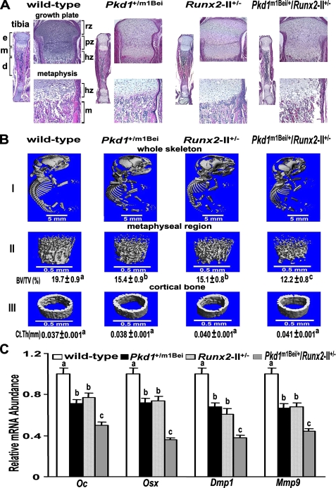
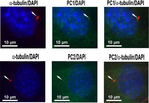
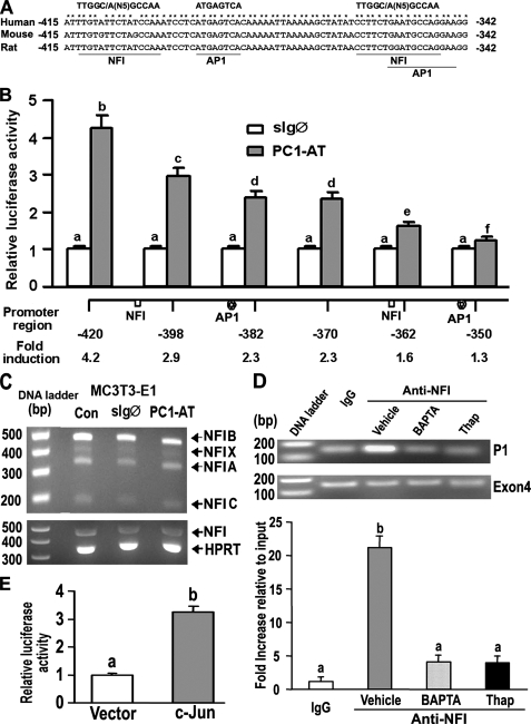
Polycystin-1 (PC1) may play an important role in skeletogenesis through regulation of the bone-specific transcription factor Runx2-II. In the current study we found that PC1 co-localizes with the calcium channel polycystin-2 (PC2) in primary cilia of MC3T3-E1 osteoblasts. To establish the role of Runx2-II in mediating PC1 effects on bone, we crossed heterozygous Pkd1(m1Bei) and Runx2-II mice to create double heterozygous mice (Pkd1(+/m1Bei)/Runx2-II(+/-)) deficient in both PC1 and Runx2-II. Pkd1(+/m1Bei)/Runx2-II(+/-) mice exhibited additive reductions in Runx2-II expression that was associated with impaired endochondral bone development, defective osteoblast-mediated bone formation, and osteopenia. In addition, we found that basal intracellular calcium levels were reduced in homozygous Pkd1(m1Bei) osteoblasts. In contrast, overexpression of a PC1 C-tail construct increased intracellular calcium and selectively stimulated Runx2-II P1 promoter activity in osteoblasts through a calcium-dependent mechanism. Site-directed mutagenesis of critical amino acids in the coiled-coil domain of PC1 required for coupling to PC2 abolished PC1-mediated Runx2-II P1 promoter activity. Additional promoter analysis mapped the PC1-responsive region to the "osteoblast-specific" enhancer element between -420 and -350 bp that contains NFI and AP-1 binding sites. Chromatin immunoprecipitation assays confirmed the calcium-dependent binding of NFI to this region. These findings indicate that PC1 regulates osteoblast function through intracellular calcium-dependent control of Runx2-II expression. The overall function of the primary cilium-polycystin complex may be to sense and transduce environmental clues into signals regulating osteoblast differentiation and bone development.


Figures

FIGURE 1.
FIGURE 1.
Combined effect of Pkd1 and Runx2-II deficiency on Runx2 isoform message expression in newborn mice. An additive reduction of Runx2-II, but not Runx2-I, message level was observed in double Pkd1+/m1Bei/Runx2-II+/- mice compared with either single heterozygous Pkd1+/m1Bei or Runx2-II+/- mice by real-time RT-PCR. Data are expressed as the mean ± S.E. from four to five individual newborn mice. Values sharing the same superscript (a, b, or c) are not significantly different at p < 0.05.
FIGURE 2.
FIGURE 2.
Additive effect of Pkd1 and Runx2-II deficiency on embryonic bone development. A, gross appearance and body weight of newborn mice. Body weight (expressed in mg) represents the mean ± S.E. from four to five individual newborn mice per genotype. Values sharing the same superscript (a or b) are not significantly different at p < 0.05. B, Alizarin red/Alcian blue staining of wild-type, Runx2-II+/-, Pkd1+/m1Bei, and double heterozygous Pkd1+/m1Bei/Runx2-II+/- newborn mice. Calcified tissues are stained red, and cartilage is stained blue. Panels I-V, respectively, represents a superior view of the skull, clavicles, hyoid bone, sternum and ribs, scapula and forelimb (left image), and caudal vertebrae (right image). Arrows indicated delayed ossification of specific bones.
FIGURE 3.
FIGURE 3.
Skeletal abnormalities in combined Pkd1- and Runx2-II-deficient newborn mice. A, hematoxylin and eosin staining of decalcified tibias. Low magnification (20×) of entire tibia and high magnification of growth plate (100×) and metaphysic (200×) shows less trabecular bone volume, a narrow bone collar, and a reduced size of the bone marrow cavity in double Pkd1+/m1Bei/Runx2-II+/- mice. e, epiphysis; m, metaphysis; d, diaphysis; rz, chondrocyte resting zone; pz, chondrocyte proliferating zone; hz, chondrocyte hypertrophic zone. B, representative three-dimensional μCT images of the complete skeleton (panel I), metaphyseal region (panel II), and cortical bone (panel III) in Pkd1+/m1Bei/Runx2-II+/- mice. Pkd1+/m1Bei/Runx2-II+/- mice exhibited wider anterior fontanelles, diminished metaphyseal bone volume, and relative preservation of cortical bone width. C, effect of Pkd1 and Runx2-II deficiency on Runx2-dependent message expression in bone by real-time RT-PCR. Double Pkd1+/m1Bei/Runx2-II+/- mice exhibited an additive reduction in osteocalcin (Oc), osterix (Osx), Dmp1, and Mmp9 compared with single heterozygous Pkd1m1Bei and Runx2-II mice. Data are expressed as the mean ± S.E. from four to five individual samples. BV/TV, bone volume/total volume; Ct.Th, cortical bone thickness. Values sharing the same superscript (a, b, or c) are not significantly different at p < 0.05.
FIGURE 4.
FIGURE 4.
Effects of combined Pkd1 and Runx2-II deficiency on bone in 6-week-old mice. A, gross appearance and body weight. Double heterozygous Pkd1+/m1Bei/Runx2-II+/- were smaller in size and exhibited a significant reduction in body weight compared with wild-type and single heterozygous Runx2-II+/- and Pkd1+/m1Bei mice. B, BMD of femurs. There was a greater reduction (22%) of femoral BMD in both male and female double heterozygous Pkd1+/m1Bei/Runx2-II+/- mice compared with Pkd1+/m1Bei (10%) and Runx2-II+/- (10%) littermates. C, μCT analysis of femurs. Double heterozygous Pkd1+/m1Bei/Runx2-II+/- mice demonstrated greater loss in both trabecular and cortical bone than single heterozygous mice. D, mineral apposition rate. There was a significant decrease in mineral apposition rate in single Pkd1+/m1Bei and Runx2-II+/- heterozygous mice compared with age-matched wild-type mice and an even greater reduction in double heterozygous Pkd1+/m1Bei/Runx2-II+/-mice, indicating an additive effect of Pkd1 and Runx2-II deficiency to impair osteoblast-mediated bone formation. Data are expressed as the mean ± S.E. from at least three individual samples. Values sharing the same superscript (a, b, or c) are not significantly different at p < 0.05.
FIGURE 5.
FIGURE 5.
PC1 regulates Runx2-II P1 promoter activity through intracellular calcium ([Ca2+]i) in MC3T3-E1 osteoblasts. A-C, cotransfection of various PC1 constructs with Runx2 P1 and P2 promoter-luciferase constructs in MC3T3-E1 osteoblasts. Overexpression of mouse PC1-AT (gain-of-function) significantly stimulated Runx2 P1 promoter activity but not the P2 promoter activity in osteoblasts (A). The control sIgØ construct had no effect. In contrast, overexpression of the PC1-ATmutant construct that disrupts coupling to PC2 failed to stimulate P1 promoter activity (B). Overexpression of human PKD1-(115-226) that also contains the coiled-coil domain stimulates Runx2 P1 but not P2 promoter activity in osteoblasts, whereas overexpression of PDK1(1-92) that lacks the coiled-coil domain fails to stimulate Runx2 P1 promoter activity in MC3T3-E1 osteoblasts (C). D and E, effect of Pkd1 on intracellular calcium ([Ca2+]i) in osteoblasts. There was a significant reduction in basal [Ca2+]i in Pkd1m1Bei/m1Bei mutant primary osteoblasts (D). Stable overexpression of the PC1-AT in MC3T3-E1 osteoblasts resulted in a significant increase in [Ca2+]i BAPTA, which chelates intracellular calcium, or thapsigargin, which depletes intracellular calcium stores, completely inhibited PC1-AT-mediated stimulation of Runx2 P1 promoter activity in osteoblasts (E). Data are expressed as the mean ± S.E. from at least three independent experiments. The asterisk indicates a significant difference from wild type and control, and values sharing the same superscript (a, b, or c) are not significantly different at p < 0.05.
FIGURE 6.
FIGURE 6.
Co-localization of PC1 and PC2 to primary cilia in osteoblasts. Co-immunostaining with α-tubulin and PC1 or PC2 antibodies was performed in MC3T3-E1 osteoblasts as described under “Experimental Procedures.” Co-localization of PC1 (upper panel) and PC2 (lower panel) to the primary cilium (upper panel) is shown. A single primary cilium (red) is shown projecting from the cell surface of an osteoblast (left). PC1 and PC2 are present in both a diffuse and localized pattern (green) in osteoblasts (middle). Co-localization of PC1 and PC2 to the primary cilium is illustrated by the orange-yellow color in the merged images (right). 4′,6-Diamidino-2-phenylindole (DAPI) blue-staining was used to identify nuclei. Images are representative samples viewed with a fluorescent microscope at a magnification of 600×.
FIGURE 7.
FIGURE 7.
NFI and AP-1 transcription factors mediate PC1-dependent activation of the P1 Runx2-II promoter. A, schematic showing two NFI and AP1 binding sites located between -415 and -342 region of the Runx2-II P1 promoter that are conserved across species. B, assessment of NF1 and AP1 function by deletion analysis of the Runx2 P1 promoter. MC3T3-E1 osteoblasts were transiently co-transfected with PC1-AT or sIgØ along with Runx2-P1 promoter-luciferase constructs that contained successive 5′ deletions. Loss of NFI and AP1 sites resulted in a progressive reduction in PC1-mediated stimulation of Runx2 P1 promoter activity. C, RT-PCR analysis of NFI-A, -B, -C, and -X isoform expression in MC3T3-E1 osteoblasts transiently transfected with either the control sIgØ or the PC1-AT constructs for 48 h. Neither PC1-AT nor sIgØ increased NFI message levels above untransfected controls (CON) in MC3T3-E1, raising the possibility that PC1 regulates NFI through post-translational modifications. HPRT, hypoxanthine-guanine phosphoribosyltransferase. D, quantitative ChIP analyses demonstrating specific interaction of NFI with Runx2-II P1 promoter. Quantitative real-time PCR was performed using set of primers indicated under “Experimental Procedures.” The upper panel is a representative ethidium bromide gel of PCR products, and the lower panel is bar graph representing mean ± S.E. from at least three independent experiments. Values are shown as relative -fold of enrichment of the promoter sequence normalized for coding region sequence versus that obtained for input samples. Values sharing the same superscript (a, b, c, and d) are not significantly different at p < 0.05. Immunoprecipitations were performed with NFI antibody or with normal rabbit IgG as described under “Experimental Procedures.” E, transient co-transfection c-Jun plasmid with Runx2 P1-420-Luc (p0.42Runx2P1-Luc) stimulated Runx2 P1 promoter activity, consistent with functional AP-1 binding sites.

References

    1. Cohen, M. M., Jr. (2006) Am. J. Med. Genet. A 140 2646-2706 - PubMed
    1. Kronenberg, H. M. (2003) Nature 423 332-336 - PubMed
    1. de Crombrugghe, B., Lefebvre, V., and Nakashima, K. (2001) Curr. Opin. Cell Biol. 13 721-727 - PubMed
    1. Ducy, P., Zhang, R., Geoffroy, V., Ridall, A. L., and Karsenty, G. (1997) Cell 89 747-754 - PubMed
    1. Komori, T., Yagi, H., Nomura, S., Yamaguchi, A., Sasaki, K., Deguchi, K., Shimizu, Y., Bronson, R. T., Gao, Y. H., Inada, M., Sato, M., Okamoto, R., Kitamura, Y., Yoshiki, S., and Kishimoto, T. (1997) Cell 89 755-764 - PubMed

Publication types

MeSH terms