. 2008 May;82(10):4823-33.
doi: 10.1128/JVI.02518-07. Epub 2008 Mar 5.

A novel methyltransferase methylates Cucumber mosaic virus 1a protein and promotes systemic spread

Affiliations

A novel methyltransferase methylates Cucumber mosaic virus 1a protein and promotes systemic spread

Min Jung Kim et al. J Virol. 2008 May.

Abstract

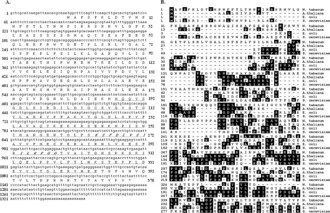
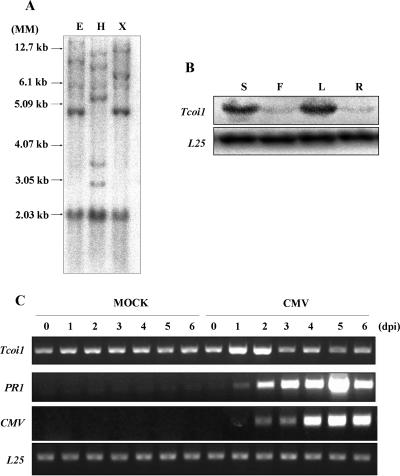
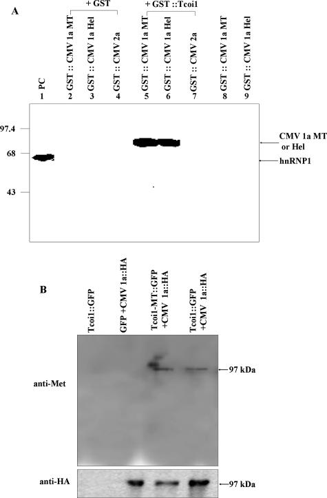
In mammalian and yeast systems, methyltransferases have been implicated in the regulation of diverse processes, such as protein-protein interactions, protein localization, signal transduction, RNA processing, and transcription. The Cucumber mosaic virus (CMV) 1a protein is essential not only for virus replication but also for movement. Using a yeast two-hybrid system with tobacco plants, we have identified a novel gene encoding a methyltransferase that interacts with the CMV 1a protein and have designated this gene Tcoi1 (tobacco CMV 1a-interacting protein 1). Tcoi1 specifically interacted with the methyltransferase domain of CMV 1a, and the expression of Tcoi1 was increased by CMV inoculation. Biochemical studies revealed that the interaction of Tcoi1 with CMV 1a protein was direct and that Tcoi1 methylated CMV 1a protein both in vitro and in vivo. The CMV 1a binding activity of Tcoi1 is in the C-terminal domain, which shows the methyltransferase activity. The overexpression of Tcoi1 enhanced the CMV infection, while the reduced expression of Tcoi1 decreased virus infectivity. These results suggest that Tcoi1 controls the propagation of CMV through an interaction with the CMV 1a protein.


Figures

FIG. 1.
FIG. 1.
Comparison of Tcoi1 nucleotide and protein sequences. (A) Nucleotide sequence of Tcoi1 cDNA and corresponding deduced amino acid sequence. The MT domain is underlined, and motif II and motif III of AdoMet-dependent MTs are marked in italics. (B) Alignment of the predicted Tcoi1 amino acid sequence with those of other UbiE-like C-MT family members. Sequences from A. thaliana (At1g23360), E. coli, and S. cerevisiae are compared. The alignments were generated from DNAStar MegAlign by using the PAM 250 table and the Jotun Hein method. The shaded residues match the consensus within two distance units.
FIG. 2.
FIG. 2.
Southern blot analysis and comparison of Tcoi1 expression patterns in various organs and in tissues from CMV-Kor- and mock-inoculated plants. (A) Tobacco genomic DNA was digested with EcoRI (E), HindIII (H), or XbaI (X), and digestion products were separated on 0.8% agarose gel. After being transferred onto a Nytran Plus membrane, the blot was hybridized with a 32P-labeled full-length Tcoi1 cDNA probe under conditions of medium stringency. Autoradiograms were visualized with a Fuji BAS 2500 phosphorimager. DNA size standards (MM) are shown at the left. (B) Accumulation of Tcoi1 transcripts in different organs. Lanes: S, stem; F, flower; L, leaf; and R, root. Tcoi1 transcripts were monitored by Northern blot analysis using the Tcoi1-specific 3′ untranslated region as a probe. The transcript level corresponding to ribosomal protein L25 was included as an internal standard for RNA quantity evaluation. (C) RT-PCR analysis of Tcoi1 genes upon CMV-Kor inoculation. Total RNA was extracted from leaf tissues at 0, 1, 2, 3, 4, 5, and 6 dpi. Tissue was isolated from the leaves of either CMV- or mock-inoculated plants. The conserved 3′ ends of CMV RNAs and PR-1 were detected as a positive control for CMV inoculation. As an internal standard for cDNA quantity evaluation, the level of L25 was monitored.
FIG. 3.
FIG. 3.
Identification of the region of Tcoi1 that is necessary for the interaction with CMV 1a protein and in vitro interaction analysis. (A) The names of the truncated constructs and the positions of amino acids (aa) are indicated at the left and above, respectively. (B) Analysis of interaction between different parts of Tcoi1 and CMV 1a protein in the yeast two-hybrid system. The interaction strength was scored by liquid assays using a sensitive luminescence detection system (Clontech). The values are the means ± standard deviations (n = 3) and are expressed as relative light units (RLU) normalized to the cell content (optical density at 600 nm [OD600]). In the evaluation of each interaction, three separate transformed colonies were assayed, and each of the assays was performed in triplicate. PC, positive control; NC, negative control. (C) In vitro GST pulldown assay of the GST-conjugated products of full-length and truncated Tcoi1 clones and in vitro-translated CMV 1a protein. [35S]Met-labeled CMV 1a was incubated with 10 μg of GST (negative control), GST-Tcoi1, GST-Tcoi1d1, GST-Tcoi1d2, GST-Tcoi1d3, or GST-Tcoi1-MT in the presence of glutathione-agarose beads. Precipitates from the binding mixture were subjected to SDS-PAGE, and proteins were visualized by autoradiography. Numbers on the left are molecular size markers.
FIG. 4.
FIG. 4.
In vivo interaction tests of CMV 1a and Tcoi1, Tcoi1d3, or Tcoi1-MT. (A) Fluorescence micrographs of Arabidopsis protoplasts coexpressing RLuc and YFP fusions. (B) BRET protein interaction analysis of Arabidopsis protoplasts. The yellow/blue ratio in living tissue after the transient coexpression of the indicated fusion proteins was measured. The letters R and Y indicate the positions of the RLuc and YFP tags, respectively. pRLuc::YFP (R-Y) and pRLuc (R) were used as the positive control and the negative control, respectively. The data were averaged from the results for three replicates. Error bars represent the standard deviations. (C and D) Arabidopsis protoplasts were cotransformed with plasmids encoding GFP and CMV 1a-HA, Tcoi1-MT-GFP and CMV 1a-HA, or Tcoi1-GFP and CMV 1a-HA. As a control, protoplasts were transformed with the plasmid encoding Tcoi1-GFP. The immunoprecipitation of the protoplast extracts was performed with anti-HA antiserum. The immunoprecipitated proteins were subjected to Western blot analysis with anti-GFP (C) or anti-HA (D) antibody.
FIG. 5.
FIG. 5.
In vitro and in vivo methylation of CMV 1a by Tcoi1. (A) Methylation of the CMV 1a MT domain and the CMV 1a Hel domain by purified Tcoi1. The protein MT assay was carried out with equal amounts (10 μg) of the indicated purified proteins in the presence of 0.25 μCi of S-adenosyl-l-[methyl-3H]methionine. Lane 1, PRMT1 and hnRNP1 (as a positive control [PC]). The reaction products were separated by SDS-10% PAGE and visualized by autoradiography. The hnRNP1 (59-kDa), GST-CMV 1a-MT (77-kDa), and GST-CMV 1a-Hel (75-kDa) bands are indicated by arrows. Molecular size markers (in kilodaltons) are indicated on the left. (B) Arabidopsis protoplasts were cotransformed with plasmids encoding the indicated proteins. As a control, protoplasts were transformed with the plasmid encoding Tcoi1-GFP. The immunoprecipitation of the protoplast extracts was performed with anti-HA antiserum. The immunoprecipitated proteins were subjected to Western blot analysis with anti-mono-/dimethylarginine (anti-Met) (upper panel) or anti-HA (lower panel) antibody.
FIG. 6.
FIG. 6.
Expression of Tcoi1 gene in transgenic tobacco plants and comparison of CMV RNA levels and patterns of CMV CP accumulation and symptom development in wild-type and transgenic plants. (A) RT-PCR comparison of the transgenic T2 tobacco plants and the untransformed control plants. As an internal standard for cDNA quantification, the level of L25 transcripts was also monitored. WT, wild type; 35S::Tcoi1-S, sense-Tcoi1 transgenic plants; 35S::Tcoi1-AS, antisense-Tcoi1 transgenic plants. (B) The levels of CMV RNA accumulation in the transgenic T2 plants, along with those in the control plants, were monitored by Northern blotting using the CMV RNA 3′ untranslated region as a probe on the indicated days after inoculation. The uninoculated upper leaves from sense-Tcoi1 and antisense-Tcoi1 plants and wild-type plants, as indicated, were monitored at 3, 5, and 7 dpi and then harvested for RNA analysis. The positions of the bands representing CMV RNAs 1, 2, 3, and 4 are shown to the right of the panel. The RNA loading control was stained with ethidium bromide (EtBr). M, molecular size marker. (C) The intensities of the bands for CMV RNA 4 on Northern blots were quantified. Boxes and error bars represent the means and standard deviations of results from three independent experiments examining relative levels of viral RNA accumulation, normalized by the intensities of the wild-type bands in the corresponding lanes. Values are expressed as log2 ratios. (D) Comparison of the CMV-Kor infection symptoms in uninoculated upper leaves of transgenic and untransformed plants at 9 dpi. The lower panels show higher magnifications of the boxed regions. (E) Comparison of CMV-Kor CP accumulation patterns in uninoculated upper leaves at 9 dpi. The accumulation of CMV CP was measured via ELISA, and the results were expressed as the absorbance at 405 nm (A405) per 45 μg of total protein (n = 30 plants). The data were analyzed by analysis of variance. Error bars indicate the standard deviations, and asterisks indicate significant differences compared with the wild-type control (P < 0.001).

Similar articles

Cited by

References

    1. Aletta, J. M., T. R. Cimato, and M. J. Ettinger. 1998. Protein methylation: a signal event in post-translational modification. Trends Biochem. Sci. 2389-91. - PubMed
    1. Ausubel, F. M., R. Brent, R. E. Kingston, D. D. Moore, J. G. Seidman, J. A. Smith, and K. Struhl. 1995. Short protocols in molecular biology. Wiley, New York, NY.
    1. Baba, S. W., G. I. Belogrudov, J. C. Lee, P. T. Lee, J. Strahan, J. N. Shepherd, and C. F. Clarke. 2004. Yeast Coq5 C-methyltransferase is required for stability of other polypeptides involved in coenzyme Q biosynthesis. J. Biol. Chem. 27910052-10059. - PubMed
    1. Borchardt, R. T., B. T. Keller, and U. Patel-Thombre. 1984. Neplanocin A. A potent inhibitor of S-adenosylhomocysteine hydrolase and of vaccinia virus multiplication in mouse L929 cells. J. Biol. Chem. 2594353-4358. - PubMed
    1. Boulanger, M. C., C. Liang, R. S. Russell, R. Lin, M. T. Bedford, M. A. Wainberg, and S. Richard. 2005. Methylation of Tat by PRMT6 regulates human immunodeficiency virus type 1 gene expression. J. Virol. 79124-131. - PMC - PubMed

Publication types