. 2008 May;82(10):4862-73.
doi: 10.1128/JVI.01202-07. Epub 2008 Mar 5.

Casein kinase II motif-dependent phosphorylation of human papillomavirus E7 protein promotes p130 degradation and S-phase induction in differentiated human keratinocytes

Affiliations

Casein kinase II motif-dependent phosphorylation of human papillomavirus E7 protein promotes p130 degradation and S-phase induction in differentiated human keratinocytes

Nicholas J Genovese et al. J Virol. 2008 May.

Abstract

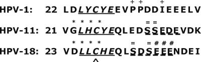
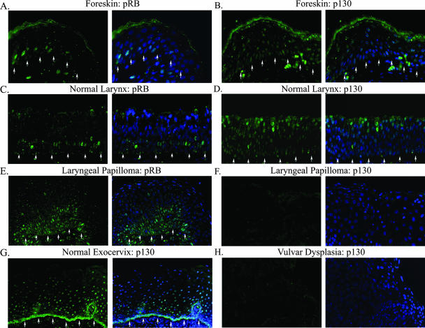
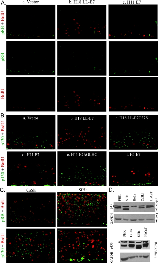
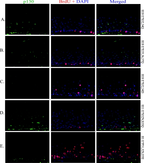
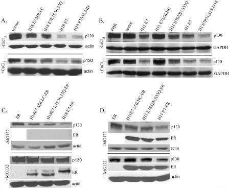
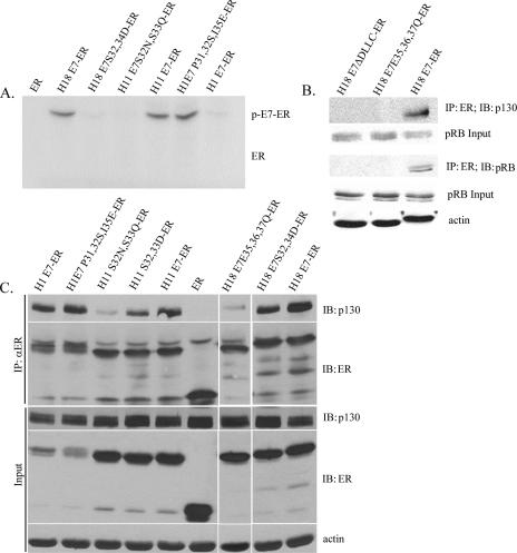
The E7 proteins of human papillomaviruses (HPVs) promote S-phase reentry in differentiated keratinocytes of the squamous epithelia to support viral DNA amplification. In this study, we showed that nuclear p130 was present in the differentiated strata of several native squamous epithelia susceptible to HPV infection. In contrast, p130 was below the level of detection in HPV-infected patient specimens. In submerged and organotypic cultures of primary human keratinocytes, the E7 proteins of the high-risk mucosotrophic HPV-18, the benign cutaneous HPV-1, and, to a lesser extent, the low-risk mucosotropic HPV-11 destabilized p130. This E7 activity depends on an intact pocket protein binding domain and a casein kinase II (CKII) phosphorylation motif. Coimmunoprecipitation experiments showed that both E7 domains were important for binding to p130 in extracts of organotypic cultures. Metabolic labeling in vivo demonstrated that E7 proteins were indeed phosphorylated in a CKII motif-dependent manner. Moreover, the efficiencies of the E7 proteins of various HPV types or mutations to induce S-phase reentry in spinous cells correlated with their relative abilities to bind and to destabilize p130. Collectively, these data support the notion that p130 controls the homeostasis of the differentiated keratinocytes and is therefore targeted by E7 for degradation to establish conditions permissive for viral DNA amplification.


Figures

FIG. 1.
FIG. 1.
Amino acid sequence alignment of E7 pocket protein binding regions of HPV-1, HPV-11, and HPV-18. Numbers represent the position of the first residue displayed. The solid underlined sequence indicates the conserved LxCxE pocket protein binding domain. The dashed underlined sequence signifies the consensus SxxD/E CKII motif. Like symbols above or below the residues denote mutated residues in mutant forms of E7 characterized in this study.
FIG. 2.
FIG. 2.
Indirect immunofluorescence analyses of pRB and p130 proteins in normal tissues and HPV-infected lesions. Shown is pRB or p130 (green) in tissue sections from formalin-fixed and paraffin-embedded neonatal foreskin (A and B), normal laryngeal tissue (C and D), normal exocervical tissue (G), HPV-11-infected laryngeal papilloma (E and F), and HPV-16-infected vulvar dysplasia (H). Each image is presented twice. The left panels reveal the pocket protein staining, whereas the right panels show merged images with DAPI-stained nuclei (blue). Arrows indicate the basal stratum. For panels F and H, the basal stratum was out of view beyond the left lower corner. All images were captured with a ×20 objective lens but are presented at slightly different magnifications for best viewing.
FIG. 3.
FIG. 3.
Simultaneous detection of BrdU and pRB or p130 in organotypic raft cultures of cervical carcinoma cell lines and in PHKs expressing wild-type E7 or E7 mutated in the LxCxE motif. PHKs were acutely transduced with recombinant retroviruses that expressed the vector alone or the indicated HPV E7 (A and B). The CaSki and SiHa cervical carcinoma cell lines (C) express HPV-16 E6 and E7. All raft cultures were exposed to BrdU for 12 h immediately prior to harvest to mark cells in S phase. All sections were imaged at ×20 magnification. (A) pRB (green) plus BrdU incorporation (red) in separate and merged images. (B) p130 (green) and BrdU incorporation (red) in merged images. (C) Raft cultures derived from CaSki and SiHa cells. Upper panels show pRB (green) and BrdU incorporation (red); lower panels show p130 (green) and BrdU (red). (D) Western blots of total cell lysates from submerged or raft cultures of PHKs of indicated cell lines to detect p130. *, a nonspecific, cross-reactive protein band.
FIG. 4.
FIG. 4.
Simultaneous detection of S-phase cells and p130 in organotypic cultures expressing E7 with gain- or loss-of-function mutations in the CKII recognition sequence or substrates. PHKs were acutely transduced with a retrovirus expressing the indicated E7 mutation. BrdU was added to the media for the last 12 h before harvest. p130 (green) and BrdU (red) incorporation are shown for substitution mutations in HPV-18 E7 (A, B, and C), HPV-11 E7 (D), or HPV-1 E7 (E). Left panels show p130 (green), middle panels show BrdU (red) plus DAPI, and right panels show merged images. All sections were imaged at ×20 magnification.
FIG. 5.
FIG. 5.
Binding and destabilization of p130 in PHKs expressing wild-type or mutated HPV E7 or E7-ER fusion proteins. PHKs were acutely transduced with a retrovirus which expressed the wild-type or mutated E7 (A and B) or E7-ER (C and D) or a control vector (empty or encoding the ER moiety) as indicated. (A and B) Lysates from subconfluent, proliferating PHK cultures (top panels) or 2 mM CaCl2-treated, confluent cultures (bottom panels) were Western blotted to reveal relative p130 levels. Actin or GAPDH was detected as a loading reference. Cultures in panel B were treated with 250 μM CHX for 6 h prior to harvest. (C and D) Western blot analyses of p130, E7-ER, and actin. The cultures were induced with 5 μM β-estradiol for 24 h prior to harvest and were further treated for 6 h with 250 μM CHX in the absence (top panels) or in the presence (bottom panels) of 50 μM MG132.
FIG. 6.
FIG. 6.
Phosphorylation of HPV E7 at the CKII motif increases binding to p130 in vivo. (A) E7 phosphorylation in vivo requires the CKII substrates. Lysates of Cos7 cells expressing ER or the indicated E7-ER fusions metabolically labeled with 32Pi were immunoprecipitated (IP) with an anti-ER (αER) antibody. The immunoprecipitates were analyzed by SDS-PAGE, and the autoradiogram is presented. “ER” indicates the expected migration of the ER moiety. “p-E7-ER” indicates the migration of the phosphorylated E7-ER fusion. (B and C) The presence of CKII substrates increases binding to p130. E7-ER was immunoprecipitated from total lysates of PHK raft cultures. IB, immunoblotting. (B) Immunoprecipitates were probed for p130 or pRB. Input lysates were probed for p130, pRB, and actin. (C) Immunoprecipitates were probed with p130 or ER antibody. Input lysates were probed with p130, ER, and actin.

References

    1. Alunni-Fabbroni, M., T. Littlewood, L. Deleu, S. Caldeira, M. Giarre, M. Dell' Orco, and M. Tommasino. 2000. Induction of S phase and apoptosis by the human papillomavirus type 16 E7 protein are separable events in immortalized rodent fibroblasts. Oncogene 192277-2285. - PubMed
    1. Baker, C. C., W. C. Phelps, V. Lindgren, M. J. Braun, M. A. Gonda, and P. M. Howley. 1987. Structural and transcriptional analysis of human papillomavirus type 16 sequences in cervical carcinoma cell lines. J. Virol. 61962-971. - PMC - PubMed
    1. Baker, G. L., M. W. Landis, and P. W. Hinds. 2005. Multiple functions of D-type cyclins can antagonize pRb-mediated suppression of proliferation. Cell Cycle 4330-338. - PubMed
    1. Banerjee, N. S., N. J. Genovese, F. Noya, W.-M. Chien, T. R. Broker, and L. T. Chow. 2006. Conditionally activated E7 proteins of high-risk and low-risk human papillomaviruses induce S phase in postmitotic, differentiated human keratinocytes. J. Virol. 806517-6524. - PMC - PubMed
    1. Barbie, D. A., B. A. Kudlow, R. Frock, J. Zhao, B. R. Johnson, N. Dyson, E. Harlow, and B. K. Kennedy. 2004. Nuclear reorganization of mammalian DNA synthesis prior to cell cycle exit. Mol. Cell. Biol. 24595-607. - PMC - PubMed

Publication types

MeSH terms

Substances

LinkOut - more resources