. 2008 Apr;135(8):1415-25.
doi: 10.1242/dev.019497. Epub 2008 Mar 5.

Sdmg1 is a conserved transmembrane protein associated with germ cell sex determination and germline-soma interactions in mice

Affiliations

Sdmg1 is a conserved transmembrane protein associated with germ cell sex determination and germline-soma interactions in mice

Diana Best et al. Development. 2008 Apr.

Abstract

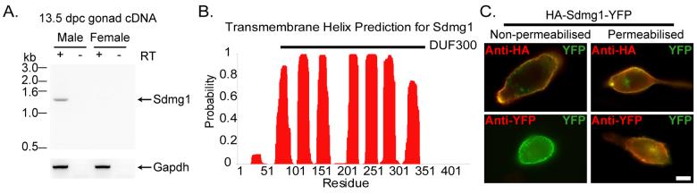
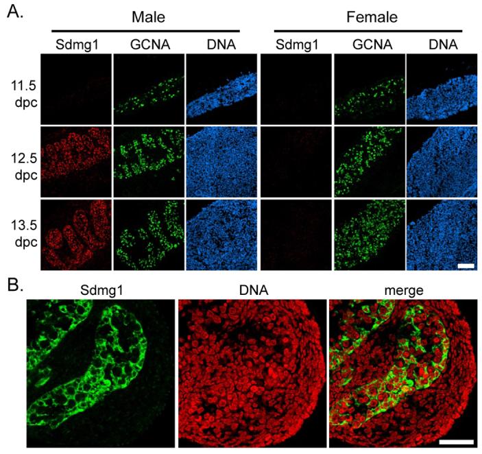
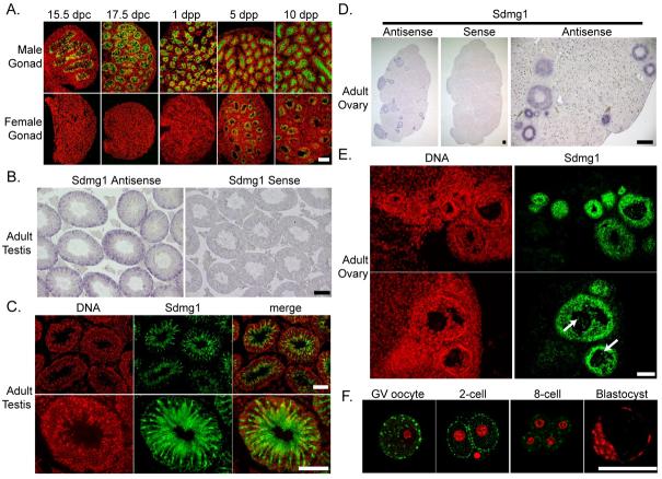
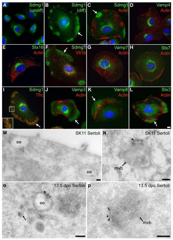
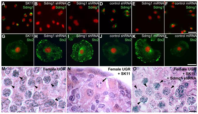
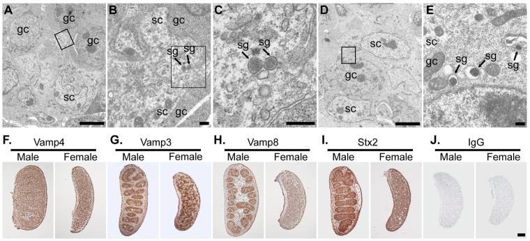
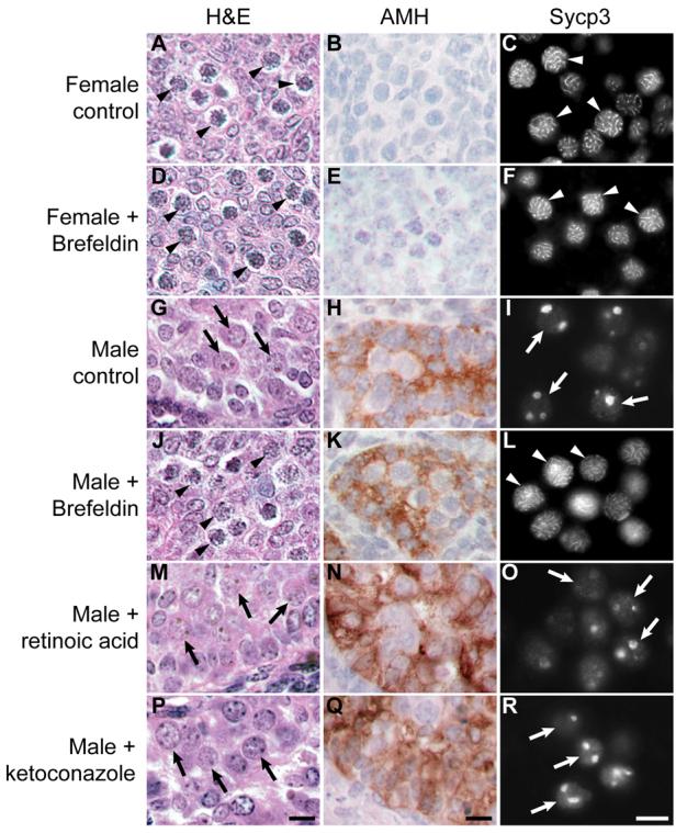
In mammals, the supporting cell lineage in an embryonic gonad communicates the sex-determining decision to various sexually dimorphic cell types in the developing embryo, including the germ cells. However, the molecular nature of the sex-determining signals that pass from the supporting cells to the germ cells is not well understood. We have identified a conserved transmembrane protein, Sdmg1, owing to its male-specific expression in mouse embryonic gonads. Sdmg1 is expressed in the Sertoli cells of embryonic testes from 12.5 dpc, and in granulosa cells of growing follicles in adult ovaries. In Sertoli cells, Sdmg1 is localised to endosomes, and knock-down of Sdmg1 in Sertoli cell lines causes mis-localisation of the secretory SNARE Stx2 and defects in membrane trafficking. Upregulation of Sdmg1 appears to be part of a larger programme of changes to membrane trafficking pathways in embryonic Sertoli cells, and perturbing secretion in male embryonic gonads in organ culture causes male-to-female germ cell sex reversal. These data suggest that changes that occur in the cell biology of embryonic Sertoli cells may facilitate the communication of male sex-determining decisions to the germ cells during embryonic development.


Figures

Figure 1
Figure 1. Sdmg1 is a Transmembrane Protein Expressed in Male Embryonic Gonads
A. RT-PCR showing male-specific expression of Sdmg1 in 13.5 dpc gonads. RT-PCR for Gapdh, and RT-PCR without reverse transcriptase (RT) are shown as controls. B. Transmembrane helix prediction for Sdmg1. The DUF300 domain is indicated with a black bar. C. Topology of Sdmg1. NIH3T3 cells were transiently transfected with HA-Sdmg1-YFP (green) and immunostained with anti-HA or anti-YFP antibodies (red). Antibody accessibility indicates that the N-terminal HA epitope is extracellular and the C-terminal YFP epitope is intracellular. Scale bar 10 μm.
Figure 2
Figure 2. Expression of Sdmg1 During Embryonic Gonad Development
A. Immunostaining for Sdmg1 (red) showing male-specific expression in the gonads from 12.5 dpc. Germ cells are labelled with anti-GCNA antibody (green), DNA is shown in blue. Scale bar 100 μm. B. Immunostaining in 13.5 dpc testes showing punctate staining for Sdmg1 (green) in Sertoli cell cytoplasm. DNA is shown in red. Scale bar 50 μm.
Figure 3
Figure 3. Expression of Sdmg1 During Gametogenesis
A. Sdmg1 is expressed in the testis cords in from 15.5 dpc to 10 dpp, and in ovarian follicles from 5 dpp. Sdmg1 immunostaining is shown in green, DNA in red. B. In situ hybridisation showing Sdmg1 mRNA expression in Sertoli cells in adult testis (antisense probe, purple precipitate). C. Immunostaining of adult testis cryosections showing Sdmg1 (green) in Sertoli cell cytoplasm. D. In situ hybridisation showing Sdmg1 mRNA expression in granulosa cells in follicles in adult ovaries. E. Immunostaining of adult ovary cryosections showing Sdmg1 (green) in the granulosa cells of follicles. Some Sdmg1 is also present in the oocyte cytoplasm (arrows). F. Immunostaining for Sdmg1 (green) in germinal vesicle (GV) stage oocytes and pre-implantation embryos. Scale bars 100 μm.
Figure 4
Figure 4. Sdmg1 is Localised to Endosomes in Sertoli Cells
A. Immunostaining shows Sdmg1 is perinuclear in undifferentiated (undiff.) SK11 Sertoli cells grown at 33°C. B. Sdmg1 is present in perinuclear and peripheral clusters (arrow) in differentiating (diff.) SK11 Sertoli cells grown at 39.5°C for 1 day. C. The peripheral clusters (arrows) of Sdmg1 staining are localised to actin-poor parts of the cell. DH. Trans-Golgi network markers Vamp4 and Stx16, and endosomal markers Vti1b, Vamp7 and Stx7 all have a perinuclear distribution in differentiating SK11 Sertoli cells. I-L. The recycling endosome marker Tfrc, and the Vamp3, Vamp8 and Stx3 SNAREs associated with secretory granules all exhibit peripheral clusters of punctate staining (arrows) in differentiating SK11 Sertoli cells. DNA is shown in blue, scale bar 10 μm. M-P. ImmunoEM for Sdmg1 (10 nm gold particles) in differentiating SK11 Sertoli cells (M,N) and 13.5 dpc Sertoli cells (O,P). Sdmg1 is localised to the limiting membrane of early endosomes (ee) and multivesicular bodies (mvb), and is often associated with an electron-dense region of the limiting membrane (arrowhead). Sdmg1 is also present on tubulo-vesicular membranes (M, arrow in P). Scale bar 100 nm.
Figure 5
Figure 5. Sdmg1 is Required for Membrane Trafficking and Secretion in SK11 Sertoli Cells
A-F. Immunostaining of undifferentiated SK11 Sertoli cell lines shows that lines stably transfected with Sdmg1 knock-down constructs (Sdmg1 shRNA and Sdmg1 miRNA) have reduced levels of Sdmg1 protein. G-L. Immunostaining of SK11 Sertoli cells after 2 days of differentiation shows that Stx2 accumulates intracellularly in lines stably transfected with Sdmg1 knock-down constructs. DNA is shown in red. Scale bars 50 μm. M-O. Histology of 11.5 dpc female urogenital ridge tissue cultured after aggregation with or without SK11 Sertoli cell lines. Most germ cells develop into female meiotic oocytes after aggregation without SK11 cells (M, arrowheads), into male prospermatogonia (N, arrow) after aggregation with SK11 cells, and into female meiotic oocytes after aggregation with Sdmg1 knock-down SK11 cells (O, arrowheads). Scale bar 10 μm.
Figure 6
Figure 6. Embryonic Sertoli Cells Are Specialised Secretory Cells
EM of 13.5 dpc testes showing secretory granules in Sertoli cells and germ cells (A-E). A,D. Low magnification image showing arrangement of germ cells (gc) and Sertoli cells (sc). B,C,E. Higher magnification of boxed regions in A, B and D respectively. Secretory granules (sg) are indicated. Scale bars 5 μm in A,D; 0.1 μm in B,C,E. F-I. Immunohistochemistry for SNAREs (brown precipitate) in 13.5 dpc ovaries and testes. F. Vamp4 (trans-Golgi network) is ubiquitously expressed. G. Vamp3 (secretory granules, endosomes) is abundant in a subset of cells in male and female gonads. H, I.Vamp8 (secretory granules, endosomes) and Stx2 (apical plasma membrane) are abundant in testis cords. J. Non-specific IgG controls show no significant staining. Scale bar 100 μm.
Figure 7
Figure 7. Perturbing Secretion Induces Male-to-Female Germ Cell Sex Reversal in Embryonic Gonad Cultures
11.5 dpc urogenital ridges were transiently treated with or without brefeldin A to block secretion, or were cultured continuously with retinoic acid or ketoconazole. Urogenital ridges were cultured for 4 days and analysed by histology (H&E) to assess germ cell development, and immunostained for anti-Mullerian Hormone (AMH, brown precipitate) to assess Sertoli cell development. Urogenital ridges were also cultured for 6 days and immunostained for Sycp3. Sycp3 labels the thread-like synaptonemal complex in meiotic oocytes, and is present as large aggregates in prospermatogonia. Examples of meiotic oocytes are labelled with arrowheads; examples of prospermatogonia are labelled with arrows. Scale bars 10 μm.

References

    1. Adams IR, McLaren A. Sexually dimorphic development of mouse primordial germ cells: switching from oogenesis to spermatogenesis. Development. 2002;129:1155–1164. - PubMed
    1. Albrecht K, Eicher E. Evidence that Sry is expressed in pre-Sertoli cells and Sertoli and granulosa cells have a common precursor. Dev. Biol. 2001;240:92–107. - PubMed
    1. Ang AL, Taguchi T, Francis S, Folsch H, Murrells LJ, Pypaert M, Warren G, Mellman I. Recycling endosomes can serve as intermediates during transport from the Golgi to the plasma membrane of MDCK cells. J. Cell Biol. 2004;167:531–543. - PMC - PubMed
    1. Arney KL, Bao S, Bannister AJ, Kouzarides T, Surani MA. Histone methylation defines epigenetic asymmetry in the mouse zygote. Int J Dev Biol. 2002;46:317–20. - PubMed
    1. Band AM, Kuismanen E. Localization of plasma membrane t-SNAREs syntaxin 2 and 3 in intracellular compartments. BMC Cell Biology. 2005;6:26. - PMC - PubMed

Publication types

MeSH terms